
Basic Mechanism of Homeostasis – Receptors, Control Centre & Effectors | Class 12 Biology
🟢 13.1.1 Basic Mechanism of Homeostasis
Every homeostatic mechanism works through three important components that continuously communicate with each other. These components are:
1️⃣ Receptors (Sensors) 2️⃣ Control Centre 3️⃣ Effectors
Together, they detect a change, make a decision, and produce the correct response to restore normal body conditions.
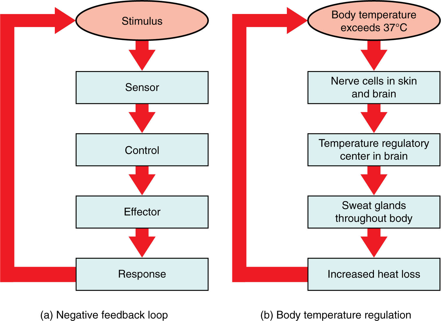
Figure 13.4 — Basic Mechanism of Homeostasis Caption: The three essential components of every homeostatic mechanism – Stimulus → Sensor → Control → Effector → Response. Attribution: OpenStax / Oregon State University Open Textbook (Educational use).
🔷 Homeostasis Flowchart
External/Internal Change
        │
        ▼
   Receptors
(Sense the change)
        │
        ▼
 Control Centre
 (Makes decision)
        │
        ▼
   Effectors
(Perform action)
        │
        ▼
 Normal Internal
 Environment Restored
🔵 1. RECEPTORS (Sensors)
📖 Definition
A Receptor (a specialized cell, tissue or organ that detects changes) is the first component of a homeostatic mechanism. Its job is to constantly monitor the body’s internal and external environment. Whenever something changes, receptors immediately detect it.
💡 Easy Explanation
Think of receptors as the security cameras of your body. They never solve the problem. They only detect the problem and send information to the control centre.
🧠 Difficult Terms
- Stimulus (any change in the environment that produces a response)
- Thermoreceptors (temperature-sensing receptors)
- Baroreceptors (pressure-sensing receptors)
- Photoreceptors (light-detecting cells found in the eye)
Examples of Receptors
| Receptor | Detects |
|---|---|
| Thermoreceptors | Temperature |
| Baroreceptors | Blood pressure |
| Chemoreceptors | Chemicals (O₂, CO₂, pH) |
| Pain receptors (Nociceptors) | Pain |
| Photoreceptors | Light |

Figure 13.5 — Thermoreceptors in Human Skin Caption: Thermoreceptors in the skin detect heat and cold and send signals to the brain. Attribution: Educational illustration (Dreamstime / similar open educational style).
Example Suppose the weather becomes very hot. ↓ Thermoreceptors in the skin detect the increase in temperature. ↓ They immediately send nerve impulses to the brain.
📦 Remember Receptors DETECT. They never make decisions.
🔵 2. CONTROL CENTRE
📖 Definition
The Control Centre (the decision-making centre of the body) receives information from receptors. It compares the received information with the Set Point (the normal value that the body tries to maintain) and decides what should happen next.
💡 Easy Explanation
The control centre works like the principal of a school. Teachers report a problem. The principal decides what action should be taken.
Main Control Centres ✔ Brain ✔ Spinal Cord ✔ Endocrine Glands (glands that produce hormones)
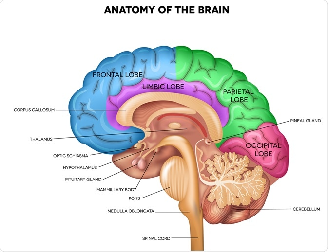
Figure 13.6 — Human Brain Showing the Hypothalamus Caption: The hypothalamus is a small but vital region of the brain that acts as the body’s thermostat. Attribution: News-Medical / educational anatomy diagram.
The Hypothalamus
The Hypothalamus (a small region of the brain that controls body temperature, hunger, thirst and many hormones) is called the Body’s Thermostat because it constantly monitors body temperature.
Set Point
A Set Point (the desired normal value maintained by the body) is the ideal condition for normal body function.
Examples
- Normal body temperature → 37°C
- Normal blood pH → 7.35–7.45
📦 Remember The Control Centre THINKS. It receives information. It makes decisions. It sends instructions.
🟠 3. EFFECTORS
📖 Definition
Effectors (muscles or glands that perform the response) receive instructions from the control centre and carry out the required action.
Examples ✔ Sweat glands ✔ Skeletal muscles ✔ Blood vessels ✔ Salivary glands

Figure 13.7 — Sweat Glands in Human Skin Caption: Cross-section of human skin showing eccrine sweat glands that help cool the body. Attribution: Lumen Learning / OpenStax style educational diagram.
Example 1 If body temperature becomes too high ↓ Hypothalamus sends signals ↓ Sweat glands become active ↓ Sweat evaporates ↓ Body cools down
Example 2 When the body becomes cold ↓ Brain sends signals ↓ Muscles begin shivering ↓ Heat is produced ↓ Body temperature rises
📦 Remember Effectors DO THE WORK. They perform the response.
🧠 Memory Trick
Remember this sentence: “Receptors Report, Control Centre Commands, Effectors Execute.”
📊 Comparison Table
| Feature | Receptors | Control Centre | Effectors |
|---|---|---|---|
| Function | Detect changes | Makes decisions | Produces response |
| Receives Signals From | Environment | Receptors | Control Centre |
| Sends Signals To | Control Centre | Effectors | Does not send |
| Examples | Thermoreceptors, Baroreceptors | Brain, Hypothalamus, Endocrine glands | Sweat glands, Muscles, Blood vessels |
🎯 Real-Life Example
Imagine a classroom: 👀 CCTV Camera = Receptor 👨🏫 Principal = Control Centre 👮 Security Guard = Effector
If a problem occurs, the camera detects it, the principal decides what to do, and the security guard takes action. Homeostasis works in exactly the same way.
⭐ Board Exam Tip
Students often confuse Receptors and Effectors.
| Receptors | Effectors |
|---|---|
| Detect change | Respond to change |
| First step | Final step |
| Sense stimulus | Carry out action |
❓ Quick Self-Assessment
- What are the three components of homeostasis?
- Why is the hypothalamus called the body’s thermostat?
- Differentiate between a receptor and an effector.
- Give two examples of receptors.
- What is meant by a “set point”?


.jpg)





0 Comments