
Photosystem 1 and 2 Explained: P680 vs P700, Z-Scheme, Light Reactions & Latest AP Biology Labs
Article Outline (for SEO & Reader Navigation)
- Introduction – Strong hook + why USA students search “photosystem 1 and 2”
- What Are Photosystem 1 and 2? – Definition & location
- History of Discovery of Photosystem 1 and 2 – Emerson, Duysens, Hill & Bendall
- Structure of Photosystem 1 and 2 – Antenna complex, reaction center, core proteins
- Photosystem 1 vs Photosystem 2 – Detailed Comparison Table
- How Photosystem 1 and 2 Work Together – The Z-Scheme
- Role of Chlorophyll a and b in Photosystem 1 and 2
- Latest Practical Work: Trending AP Biology Labs on Photosystem 1 and 2 (2025) – Floating leaf disk, DPIP reduction, chlorophyll fluorescence imaging
- Common Exam Tips & Mistakes for AP Biology Students
- FAQ – Photosystem 1 and 2
- Conclusion + CTA
Introduction
Every AP Biology student in the USA searches “photosystem 1 and 2” during light reactions revision. These two massive protein-pigment complexes in the thylakoid membrane are the heart of the light-dependent reactions. They convert sunlight into ATP and NADPH — the energy currency that powers the entire planet.

What Are Photosystem 1 and 2?
Photosystem 1 and 2 are large multi-subunit protein complexes embedded in the thylakoid membrane of chloroplasts. They contain hundreds of pigment molecules (mainly chlorophyll a and b plus carotenoids) organized into light-harvesting antenna complexes and reaction centers.
- Photosystem 2 (PSII) starts the electron flow by splitting water.
- Photosystem 1 (PSI) finishes it by reducing NADP⁺ to NADPH.
Both are named based on discovery order, not function — a fact that confuses thousands of students every year.
History of Discovery of Photosystem 1 and 2
The story of photosystem 1 and 2 begins in the 1940s–1960s with American scientist Robert Emerson.
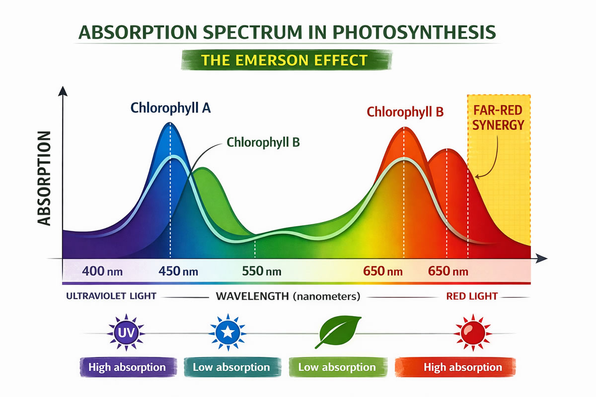
1943 – The Red Drop Effect Emerson found that photosynthesis efficiency drops sharply above 680 nm even though chlorophyll a still absorbs some far-red light.
1957 – Emerson Enhancement Effect When short red light (<680 nm) and far-red light (>680 nm) were given together, the rate of oxygen evolution doubled. This proved two cooperating light reactions exist.
1960 – Z-Scheme Proposed Robert Hill and Fay Bendall (UK) published the Z-scheme model linking the two photosystems in series. Louis Duysens (Netherlands) formally named them Photosystem I and Photosystem II based on discovery order (PSI discovered first).
1969 – H.T. Witt isolated P680 (PSII reaction center). P700 (PSI) had already been identified by Bessel Kok in 1956.
This historical numbering is why photosystem 1 and 2 act in the opposite order of their names — a classic AP Biology exam trick question.
Structure of Photosystem 1 and 2
Both photosystem 1 and 2 share the same basic architecture:
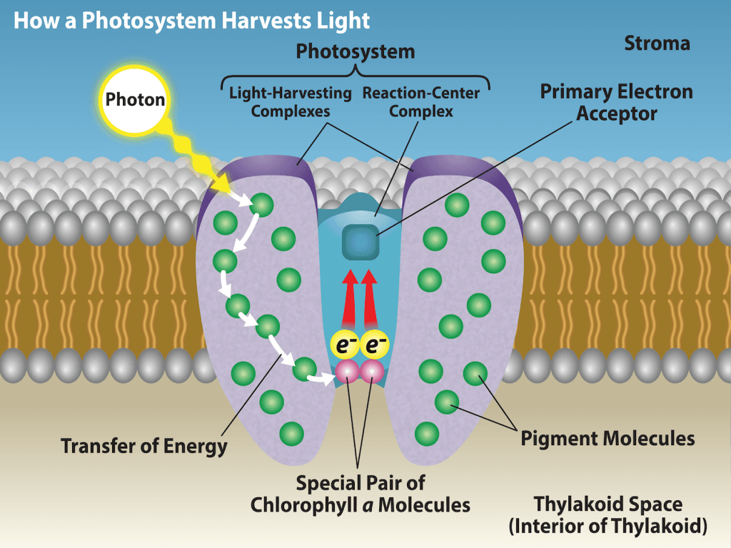
1. Antenna Complex (Light-Harvesting Complex) Hundreds of chlorophyll a, chlorophyll b, and carotenoid molecules arranged like a funnel. They capture photons and transfer energy via resonance energy transfer (Förster mechanism) to the reaction center with ~95–100% efficiency.

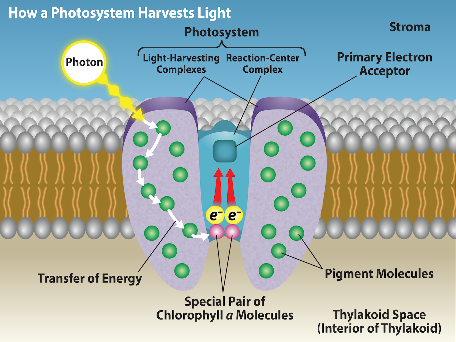
2. Reaction Center
- Photosystem 2: Special pair of chlorophyll a = P680 (absorbs best at 680 nm).
- Photosystem 1: Special pair of chlorophyll a = P700 (absorbs best at 700 nm).
3. Core Proteins & Electron Carriers PSII contains the Oxygen-Evolving Complex (OEC) with a Mn₄CaO₅ cluster. PSI has iron-sulfur centers that pass electrons to ferredoxin.
Photosystem 1 vs Photosystem 2 – Detailed Comparison Table
| Feature | Photosystem 2 (PSII) | Photosystem 1 (PSI) |
|---|---|---|
| Reaction Center | P680 | P700 |
| Primary Function | Water splitting + O₂ release | NADP⁺ → NADPH |
| Location in Thylakoid | Grana stacks | Stroma lamellae |
| Electron Source | H₂O (via OEC) | From PSII via ETC |
| Electron Acceptor | Pheophytin | A₀ (special chlorophyll) |
| Size | Larger (~20+ subunits) | Slightly smaller |
| Pigments | ~250 chlorophylls + carotenoids | ~100–200 chlorophylls + carotenoids |
How Photosystem 1 and 2 Work Together – The Z-Scheme
Photosystem 1 and 2 operate in series in the non-cyclic electron flow (Z-scheme).
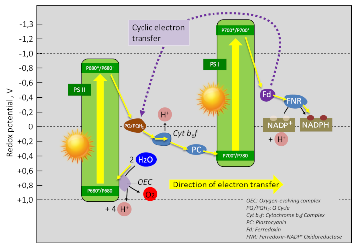
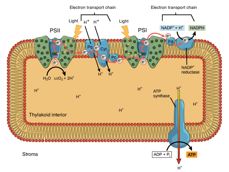
- Light excites P680 in PSII → electrons lost → replaced by splitting water (photolysis).
- Electrons flow through plastoquinone → cytochrome b₆f → plastocyanin.
- Light re-energizes electrons at P700 in PSI → electrons reduce NADP⁺ to NADPH.
- Proton gradient drives ATP synthase.
Cyclic flow (only PSI) produces extra ATP when the plant needs more energy than reducing power.
Latest Practical Work: Trending AP Biology Labs on Photosystem 1 and 2 (2025)
USA students and teachers now focus on these high-engagement labs that directly demonstrate photosystem 1 and 2 activity:
1. Floating Leaf Disk Assay (Most Popular 2025 Lab) Students punch spinach disks, infiltrate with sodium bicarbonate, and count floating disks under light. Measures net photosynthetic rate and directly links to PSII oxygen production.


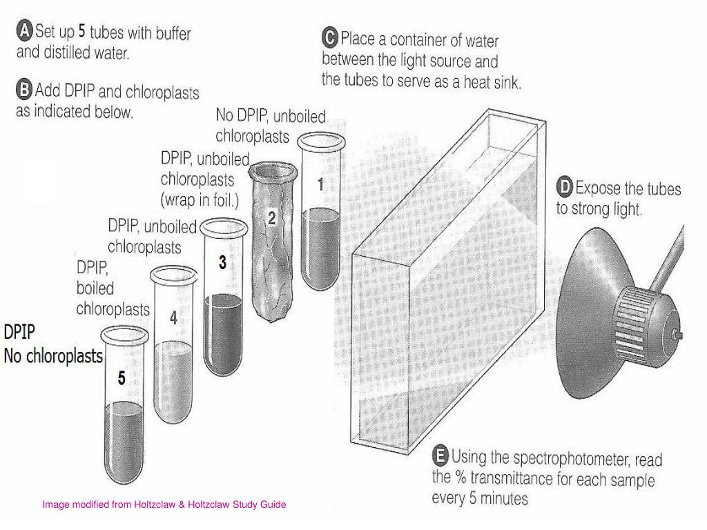
2. DPIP Reduction Lab (Classic but Still Trending) DPIP (blue) is reduced to colorless by electrons from PSII. Students measure absorbance over time to quantify electron flow through photosystem 1 and 2.
3. Chlorophyll Fluorescence Imaging (New 2024–2025 Trend) Using handheld fluorometers or phone apps, students measure Fv/Fm ratio to assess photosystem health under stress — a real-world application used in research labs.
FAQ – Photosystem 1 and 2
Q1. Why is Photosystem 2 called “2” if it acts first? It was discovered second. The numbering is historical, not functional.
Q2. What is the difference between P680 and P700? P680 (PSII) has higher redox potential and splits water. P700 (PSI) has lower potential and reduces NADP⁺.
Q3. Where are photosystem 1 and 2 located? Both in thylakoid membrane — PSII in grana, PSI in stroma lamellae.
Q4. What is the Z-scheme? The zigzag electron pathway from water → PSII → PSI → NADP⁺.
Q5. Why do we use spinach in labs? High chlorophyll content and easy pigment extraction.


.jpg)


+by+Blacklotus+Landscaping.jpg)


0 Comments