
MDCAT Biology Diagram Mistakes 2026: 50 Diagrams Students Misread in the Exam

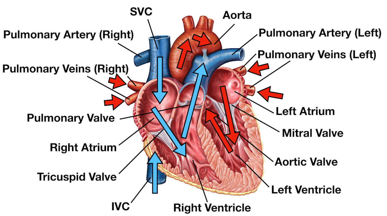
Image source: Heart Blood Flow diagram from Simplico.org (educational resource). Used for illustrative/educational purposes in this guide. Please verify current licensing and provide appropriate attribution from the original source when republishing.
Diagram 1: Pulmonary artery vs pulmonary vein
Common Mistake: Students know pulmonary artery carries deoxygenated blood but become confused when diagrams include pressure or wall thickness clues that appear to contradict the general “artery = thick wall + high pressure” rule memorised for systemic circulation.
Correct Concept: Pulmonary artery carries deoxygenated blood from the right ventricle to the lungs. It normally has thinner walls and lower pressure than the aorta. In pulmonary hypertension the pressure rises but the blood remains deoxygenated until it reaches the lungs.
Why Students Get Confused: They over-generalise systemic circulation rules and fail to apply the pulmonary exception when extra visual clues such as pressure annotations or wall thickness are present in the diagram.
Tricky MDCAT MCQ: A labelled diagram of the heart shows a vessel originating from the right ventricle with thinner walls than the vessel leaving the left ventricle. The blood in this vessel appears darker and the diagram indicates moderately elevated pressure within it. Which statement provides the most accurate interpretation?
A. The vessel is the aorta and the darker colour results from deoxygenated blood during intense exercise. B. The vessel is the pulmonary artery carrying deoxygenated blood; the thinner wall and pressure values are consistent with the pulmonary circuit although elevated pressure may indicate pulmonary hypertension. C. The vessel must be the pulmonary vein because darker colour indicates oxygenated blood returning to the heart. D. The thinner wall confirms it is a vein and the origin from the right ventricle represents a labelling error in the diagram.
Answer: B
Explanation: The vessel originates from the right ventricle and therefore must be the pulmonary artery. It carries deoxygenated blood shown by darker colour. Pulmonary arteries normally have thinner walls and lower pressure than the aorta. Moderately elevated pressure here suggests possible pulmonary hypertension but does not change the identity of the vessel or the type of blood it carries.
Quick Exam Tip: Always prioritise vessel origin from the right ventricle plus darker blood colour plus direction toward lungs when identifying the pulmonary artery, regardless of any pressure or wall thickness clues shown in the diagram.
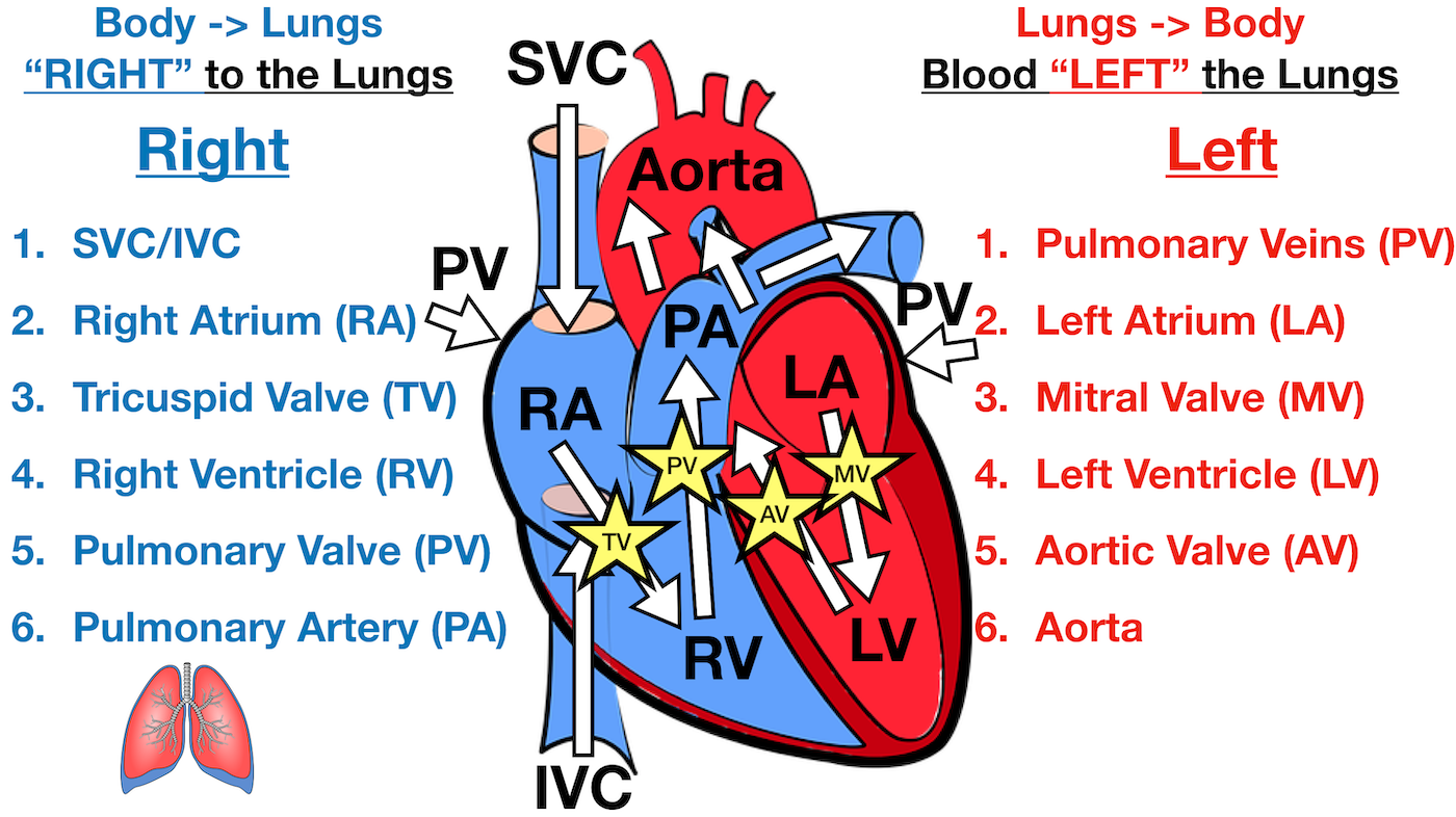
Image source: Heart Blood Flow diagram from Simplico.org (educational resource). Used for illustrative/educational purposes. Verify licensing and attribute from original source.
Diagram 2: Right side vs left side of heart
Common Mistake: Students correctly identify the sides but struggle when questions involve clinical consequences of failure on one side or when diagrams include fetal circulation shunts.
Correct Concept: The right side receives deoxygenated blood via vena cava and pumps it to the lungs through the pulmonary circuit at lower pressure. The left side receives oxygenated blood from the lungs and pumps it to the body through the systemic circuit at higher pressure. The septum prevents mixing of blood.
Why Students Get Confused: They memorise the basic rule but fail to connect side-specific failure with distinct circulatory consequences when extra clinical or pressure information appears in the diagram.
Tricky MDCAT MCQ: A heart diagram shows the right ventricular wall thinner than the left ventricular wall with blood from the right ventricle directed toward the lungs. If the right ventricle fails while the left ventricle continues to function normally, which of the following is the most likely immediate consequence?
A. Pulmonary edema due to blood backing up into the pulmonary veins. B. Systemic venous congestion and peripheral edema due to blood backing up into the superior and inferior vena cava. C. Immediate and severe drop in systemic arterial pressure because oxygenated blood cannot reach the tissues. D. No significant effect on either circuit because the left ventricle can fully compensate.
Answer: B
Explanation: Right ventricular failure causes backup of blood into the systemic venous system (vena cava), leading to systemic venous congestion and peripheral edema. Pulmonary edema occurs when the left ventricle fails. The left ventricle continues to pump oxygenated blood to the body, so systemic arterial pressure does not collapse immediately.
Quick Exam Tip: Right heart failure produces systemic backup (raised JVP, leg swelling). Left heart failure produces pulmonary backup (dyspnea, pulmonary edema). Always link the failing side to its circuit and then to the resulting clinical picture.
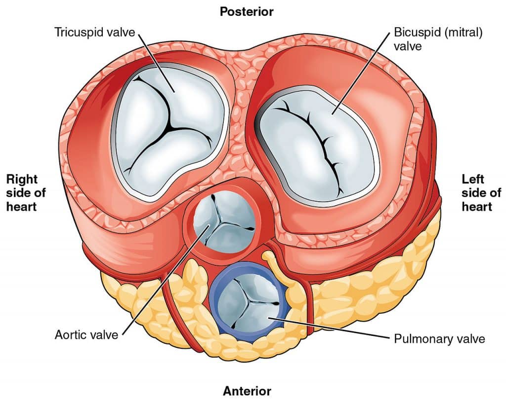
Image source: TeachMeAnatomy – The Heart Valves (educational anatomy resource). Used for illustrative/educational purposes. Verify licensing and attribute from original source.
Diagram 3: Bicuspid valve vs tricuspid valve
Common Mistake: Students remember the number of cusps but mix up clinical consequences such as the effect of stenosis on the pulmonary versus systemic circuit.
Correct Concept: Tricuspid valve has three cusps and lies between the right atrium and right ventricle. Bicuspid (mitral) valve has two cusps and lies between the left atrium and left ventricle. Both are atrioventricular valves that prevent backflow during ventricular contraction.
Why Students Get Confused: The similar names and close anatomical positions lead to swapping of cusp numbers and clinical implications when questions combine structure with consequence.
Tricky MDCAT MCQ: A diagram shows a valve with three triangular cusps located between the right atrium and right ventricle. If this valve becomes stenotic, which of the following is the most direct consequence?
A. Reduced blood flow from left atrium to left ventricle leading to pulmonary congestion. B. Reduced blood flow from right atrium to right ventricle leading to systemic venous congestion. C. Increased pressure in the aorta due to backflow from the left ventricle. D. Immediate failure of the pulmonary semilunar valve.
Answer: B
Explanation: Stenosis of the tricuspid valve obstructs blood flow from right atrium to right ventricle. This causes backup into the systemic veins, producing systemic venous congestion. The left side and aorta are not directly affected by tricuspid stenosis.
Quick Exam Tip: Tricuspid problems affect the right side and systemic venous return. Bicuspid (mitral) problems affect the left side and pulmonary venous return. Remember “Tri = Right circuit”.
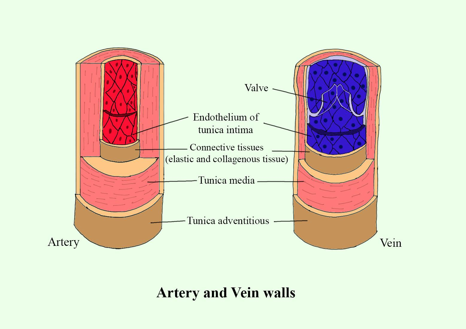
Image source: Vedantu / educational biology resource (labeled artery and vein walls diagram). Used for illustrative/educational purposes. Verify licensing and attribute from original source.
Diagram 4: Artery vs vein vs capillary
Common Mistake: Students apply the general rule that arteries carry oxygenated blood without considering the pulmonary artery exception when diagrams show mixed systemic and pulmonary vessels.
Correct Concept: Arteries carry blood away from the heart and generally have thick walls. Veins carry blood toward the heart, have thinner walls and contain valves. Capillaries are thin-walled exchange vessels. The pulmonary artery is the exception that carries deoxygenated blood.
Why Students Get Confused: Strong generalisation of systemic circulation rules causes errors when diagrams include the pulmonary circuit or pressure differences.
Tricky MDCAT MCQ: A diagram shows a vessel with thick muscular walls and narrow lumen carrying blood away from the heart. Another vessel in the same diagram has thinner walls, wider lumen and valves, carrying blood toward the heart. A third set of tiny vessels connects them. Which statement correctly identifies all three and notes any exception?
A. All three are systemic vessels and the first always carries oxygenated blood. B. The first is an artery (may carry oxygenated or deoxygenated blood depending on location), the second is a vein and the third are capillaries for exchange. C. The first must be the pulmonary vein because it carries blood away from the heart. D. The vessel with valves must be an artery because arteries have higher pressure.
Answer: B
Explanation: Arteries carry blood away from the heart regardless of oxygenation status. The pulmonary artery is the main exception carrying deoxygenated blood. Veins carry blood toward the heart and possess valves. Capillaries are the exchange vessels connecting arteries and veins.
Quick Exam Tip: Artery = away from heart. Vein = toward heart + valves. Always check origin and destination before assuming oxygenation status.
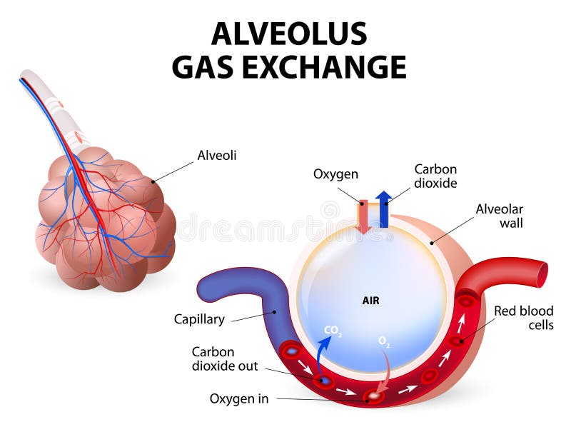
Image source: Dreamstime educational illustration – Alveolus Gas Exchange. Used for illustrative/educational purposes. Verify licensing and attribute from original source.
Diagram 5: Alveolus gas exchange direction
Common Mistake: Students reverse the direction of gas movement or fail to adjust for altered partial pressure gradients when the diagram shows a diseased lung.
Correct Concept: Oxygen diffuses from alveoli (higher partial pressure) into blood. Carbon dioxide diffuses from blood (higher partial pressure) into alveoli. The direction is driven by partial pressure gradients across the thin respiratory membrane.
Why Students Get Confused: They apply tissue-level gas exchange rules to the lungs or ignore how fibrosis or emphysema changes gradients in the diagram.
Tricky MDCAT MCQ: A diagram of an alveolus shows arrows indicating gas movement. In a patient with pulmonary fibrosis the alveolar-capillary membrane is thickened. Which change in gas exchange is most likely to be observed in the diagram?
A. Oxygen movement from blood into alveolus increases. B. Both oxygen and carbon dioxide movement slow due to increased diffusion distance. C. Carbon dioxide movement from alveolus into blood increases. D. Direction of both gases reverses completely.
Answer: B
Explanation: Thickening of the alveolar-capillary membrane increases diffusion distance and slows the rate of gas exchange for both oxygen and carbon dioxide. The direction remains the same; only the rate decreases.
Quick Exam Tip: Lungs always move oxygen in and carbon dioxide out. Disease changes rate or efficiency, not direction. Check membrane thickness and partial pressure values in the diagram.

Image source: Quizlet / Pearson Education – Structural relationship of the trachea and esophagus. Used for illustrative/educational purposes. Verify licensing and attribute from original source.
Diagram 6: Trachea vs esophagus
Common Mistake: Students confuse the anterior-posterior relationship or the presence of cartilage rings.
Correct Concept: The trachea is the anterior windpipe supported by C-shaped cartilage rings. The esophagus is the posterior food pipe with a muscular wall and no cartilage.
Why Students Get Confused: Both are longitudinal tubes in the neck and appear together in diagrams; students mix their relative positions and structural features.
Tricky MDCAT MCQ: A diagram of the neck in cross-section shows two parallel tubes. The anterior tube possesses C-shaped cartilage rings and an open lumen for air. The posterior tube has a thick muscular wall and no cartilage. Which statement correctly identifies both structures and their primary functions?
A. Anterior tube is esophagus carrying food; posterior tube is trachea carrying air. B. Anterior tube is trachea carrying air; posterior tube is esophagus carrying food. C. Both tubes are parts of the respiratory tract. D. The tube with cartilage is the esophagus because it requires structural support during swallowing.
Answer: B
Explanation: The anterior tube with C-shaped cartilage is the trachea (airway). The posterior muscular tube without cartilage is the esophagus (food pipe). Cartilage prevents collapse of the airway; the esophagus does not need it.
Quick Exam Tip: Trachea is always anterior with cartilage rings. Esophagus is always posterior and muscular. Remember “Trachea in front like a shield”.

Image source: Supercoloring.com – Inhalation and exhalation diaphragm movement diagram (educational coloring/illustration resource). Used for illustrative/educational purposes. Verify licensing and attribute from original source.
Diagram 7: Diaphragm during inhalation and exhalation
Common Mistake: Students reverse the direction of diaphragmatic movement during inhalation and exhalation.
Correct Concept: During inhalation the diaphragm contracts and flattens, moving downward and increasing thoracic volume. During exhalation the diaphragm relaxes and domes upward, decreasing thoracic volume.
Why Students Get Confused: They visualise the dome shape and incorrectly associate upward movement with inhalation.
Tricky MDCAT MCQ: A breathing diagram shows the diaphragm in a flattened position moved downward with expanded rib cage and inflated lungs. Which phase of breathing is occurring and what happens to thoracic pressure?
A. Exhalation with increased thoracic pressure. B. Inhalation with decreased thoracic pressure. C. Exhalation with decreased thoracic pressure. D. Inhalation with increased thoracic pressure.
Answer: B
Explanation: Flattened and lowered diaphragm indicates contraction during inhalation. Thoracic volume increases, pressure decreases and air rushes into the lungs.
Quick Exam Tip: Inhale = diaphragm down and flat. Exhale = diaphragm up and domed. Remember “Down for In”.
Image source: Medicine LibreTexts – Microscopic Anatomy of the Kidney (educational open resource). Used for illustrative/educational purposes. Verify licensing and attribute from original source.
Diagram 8: Bowman’s capsule vs glomerulus
Common Mistake: Students use “Bowman’s capsule” and “glomerulus” as interchangeable terms.
Correct Concept: The glomerulus is the knot of capillaries where ultrafiltration begins. Bowman’s capsule is the cup-shaped structure surrounding the glomerulus that collects the filtrate.
Why Students Get Confused: Both structures form the renal corpuscle and always appear together in nephron diagrams.
Tricky MDCAT MCQ: A nephron diagram labels a structure as the site of ultrafiltration. The structure consists of a capillary tuft surrounded by a cup-shaped capsule. Which component is primarily responsible for the initial formation of filtrate?
A. The capillary tuft (glomerulus) where blood is filtered under pressure. B. The cup-shaped capsule alone. C. The junction between afferent and efferent arterioles. D. The basement membrane only.
Answer: A
Explanation: Ultrafiltration occurs across the glomerular capillaries under hydrostatic pressure. Bowman’s capsule collects the filtrate but does not perform the filtration itself.
Quick Exam Tip: Glomerulus = filtration site (capillary ball). Bowman’s capsule = collector cup. The pair together forms the renal corpuscle.
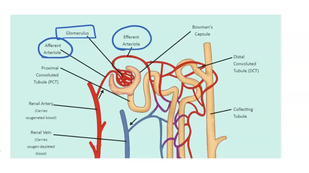
Image source: Numerade educational nephron structure diagram. Used for illustrative/educational purposes. Verify licensing and attribute from original source.
Diagram 9: PCT vs DCT
Common Mistake: Students assume both convoluted tubules perform identical reabsorption functions.
Correct Concept: The proximal convoluted tubule performs bulk obligatory reabsorption of water, glucose, amino acids and most ions. The distal convoluted tubule performs selective regulated reabsorption under the influence of aldosterone and ADH.
Why Students Get Confused: Both tubules are convoluted and located in the cortex, making them appear similar in simplified diagrams.
Tricky MDCAT MCQ: A nephron diagram shows a region with maximum reabsorption of glucose and amino acids occurring. Another region nearby shows reabsorption of sodium and water that can be increased or decreased by aldosterone and ADH. Which regions are correctly matched to these functions?
A. Proximal convoluted tubule for glucose and amino acids; distal convoluted tubule for regulated sodium and water reabsorption. B. Distal convoluted tubule for glucose and amino acids; proximal convoluted tubule for regulated sodium and water. C. Both regions perform identical functions. D. Loop of Henle performs all glucose reabsorption.
Answer: A
Explanation: The proximal convoluted tubule reabsorbs nearly all glucose and amino acids as well as the majority of water and salts. The distal convoluted tubule fine-tunes sodium and water reabsorption under hormonal control.
Quick Exam Tip: PCT = primary bulk reabsorption. DCT = regulated fine-tuning. Remember “PCT does the heavy lifting first”.
Image source: Medicine LibreTexts (same high-quality nephron resource as above, reused for consistency in this section). Used for illustrative/educational purposes. Verify licensing and attribute from original source.
Diagram 10: Loop of Henle function
Common Mistake: Students believe the Loop of Henle directly reabsorbs most of the water filtered.
Correct Concept: The Loop of Henle creates and maintains the medullary osmotic gradient through the countercurrent multiplier system. This gradient allows the collecting duct to reabsorb water under the influence of ADH, enabling production of concentrated urine.
Why Students Get Confused: They focus on water movement in the descending limb and overlook the overall gradient creation essential for the entire kidney.
Tricky MDCAT MCQ: A nephron diagram shows the Loop of Henle with arrows indicating countercurrent flow and a medullary osmolarity gradient increasing toward the papilla. If the Loop of Henle is damaged, which of the following is the most direct consequence?
A. Immediate cessation of all filtration in the glomerulus. B. Inability to create the medullary osmotic gradient, leading to production of large volumes of dilute urine. C. Complete stoppage of glucose reabsorption in the proximal tubule. D. Increased reabsorption of sodium in the distal convoluted tubule only.
Answer: B
Explanation: The countercurrent multiplier in the Loop of Henle establishes the medullary gradient necessary for water reabsorption in the collecting duct under ADH. Damage prevents concentration of urine, resulting in large volumes of dilute urine.
Quick Exam Tip: Loop of Henle = gradient builder. Collecting duct = water reabsorber using that gradient. Damage to the loop prevents urine concentration.
Diagram 11: Collecting duct vs ureter
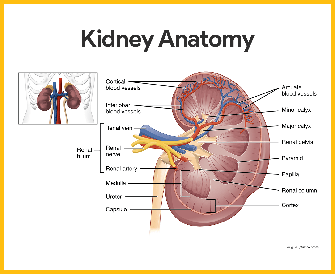
Image source: Nurseslabs – Kidney Anatomy (educational medical resource). Used for illustrative/educational purposes. Verify licensing and attribute from the original source.
Common Mistake: Students think the ureter concentrates urine or that the collecting duct only transports urine without any processing.
Correct Concept: The collecting duct is the final site of urine concentration inside the kidney (under ADH control). The ureter is a muscular tube that simply transports already-concentrated urine from the kidney to the bladder.
Why Students Get Confused: Both appear in the same excretory system diagrams and are part of the urine pathway after the nephron.
Tricky MDCAT MCQ: A diagram shows a structure inside the kidney where water reabsorption is regulated by hormones to produce concentrated urine, which then enters a thick muscular tube leading to the bladder. Which structures are correctly identified and matched to their functions?
A. Collecting duct (inside kidney, concentration) and ureter (muscular transport tube). B. Ureter (inside kidney, concentration) and collecting duct (transport tube). C. Both structures concentrate urine equally. D. The muscular tube inside the kidney is responsible for concentration.
Answer: A
Explanation: Final concentration occurs in the collecting duct inside the kidney. The ureter has no role in concentration; it only transports urine via peristalsis.
Quick Exam Tip: Collecting duct = inside kidney + concentration. Ureter = outside kidney + transport only.
Diagram 12: Kidney vs nephron

Image source: Educational anatomy resource (detailed kidney anatomy). Used for illustrative/educational purposes. Verify licensing and attribute from the original source.
Common Mistake: Students use “kidney” and “nephron” as if they are the same thing.
Correct Concept: The kidney is the organ. The nephron is the microscopic structural and functional unit (over one million per kidney) that performs filtration and urine formation.
Why Students Get Confused: Diagrams often show both the whole kidney and the detailed nephron side by side.
Tricky MDCAT MCQ: A question refers to “the structural and functional unit of the kidney” and shows a microscopic diagram containing glomerulus, Bowman’s capsule, PCT, Loop of Henle and DCT. What is being described?
A. The entire kidney. B. One nephron. C. The ureter. D. The renal pelvis.
Answer: B
Explanation: The nephron is the microscopic structural and functional unit. The kidney is the organ made up of approximately one million nephrons.
Quick Exam Tip: Kidney = organ. Nephron = tiny functional unit (millions per kidney).
Diagram 13: Xylem vs phloem
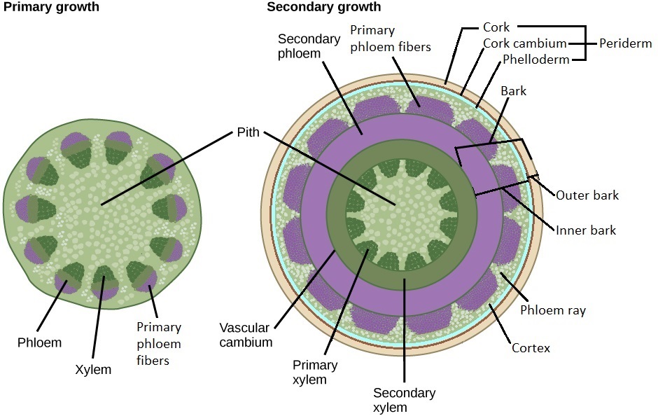
Image source: Biology LibreTexts – Secondary stem cross-section (educational open textbook). Used for illustrative/educational purposes. Verify licensing and attribute from the original source.
Common Mistake: Students mix up transport direction and whether the tissue is living or dead at maturity.
Correct Concept: Xylem transports water and minerals upward (dead cells with lignified walls). Phloem transports organic food bidirectionally (living sieve tube elements).
Why Students Get Confused: Both tissues are shown together in vascular bundles with arrows that are easy to overlook.
Tricky MDCAT MCQ: A stem cross-section diagram shows a central tissue with thick lignified walls and an outer tissue with thinner walls. Arrows show upward movement in the central tissue and movement in both directions in the outer tissue. Which identification is correct?
A. Central tissue = phloem (living, bidirectional); outer tissue = xylem (dead, upward). B. Central tissue = xylem (dead at maturity, upward water transport); outer tissue = phloem (living, bidirectional food transport). C. Both tissues transport water upward only. D. Both tissues are dead at maturity.
Answer: B
Explanation: Xylem is central in most stems, dead at maturity and transports water/minerals upward. Phloem is peripheral, living and transports food bidirectionally.
Quick Exam Tip: Xylem = upward water (dead, lignified). Phloem = food both ways (living).
Diagram 14: Stomatal pore vs guard cells
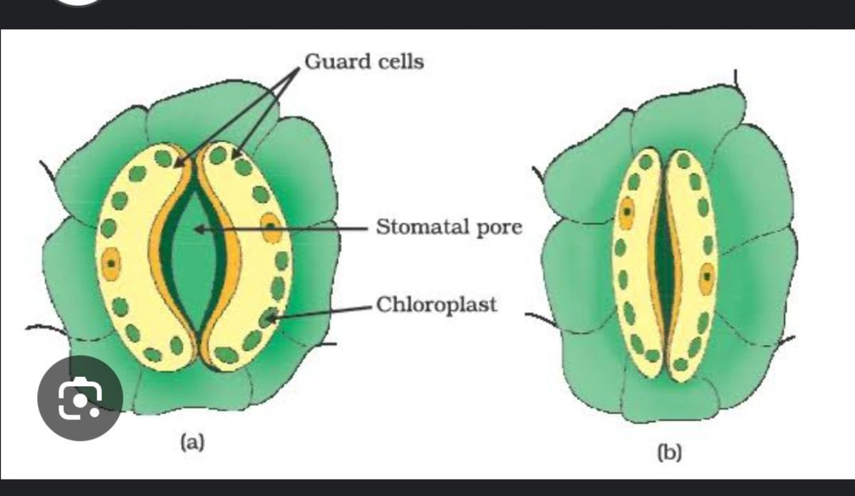
Image source: Educational biology resource – stomatal pore and guard cells. Used for illustrative/educational purposes. Verify licensing and attribute from the original source.
Common Mistake: Students think the pore opens and closes passively or that guard cells have no active role.
Correct Concept: Guard cells actively control the stomatal pore by changing turgor pressure through ion (mainly K⁺) movement and osmosis. They contain chloroplasts and have unevenly thickened walls.
Why Students Get Confused: The pore looks like a simple hole; the active physiology of guard cells is not always obvious in basic diagrams.
Tricky MDCAT MCQ: A stomatal diagram shows turgid guard cells with the pore open. Which mechanism correctly explains the opening?
A. Loss of K⁺ from guard cells causes water to leave and the pore opens. B. Entry of K⁺ into guard cells lowers water potential, water enters by osmosis, turgor rises and the pore opens. C. Thick walls alone force the pore open without ion movement. D. Passive shrinking of guard cells opens the pore.
Answer: B
Explanation: K⁺ entry lowers water potential → water enters by osmosis → turgor increases → thinner outer walls bulge outward → pore opens.
Quick Exam Tip: Turgid guard cells = open pore. Flaccid = closed. Ion movement drives the change.
Diagram 15: Palisade mesophyll vs spongy mesophyll
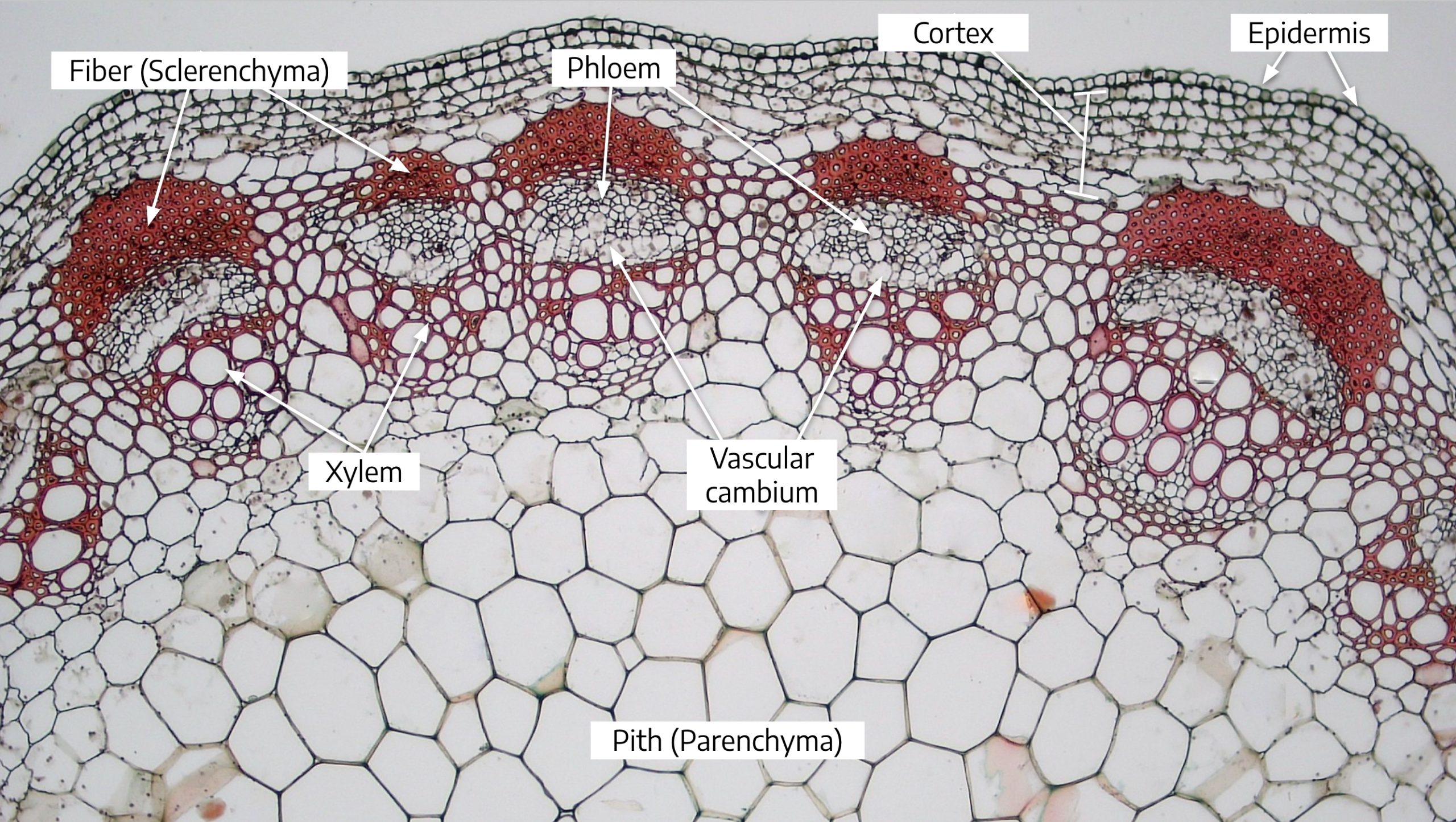
Image source: Educational plant anatomy resource – leaf/stem cross-section showing mesophyll layers. Used for illustrative/educational purposes. Verify licensing and attribute from the original source.
Common Mistake: Students assume both layers contribute equally to photosynthesis.
Correct Concept: Palisade mesophyll (upper, columnar cells, dense chloroplasts) is the main photosynthetic tissue. Spongy mesophyll (lower, irregular cells, large air spaces) facilitates gas exchange.
Why Students Get Confused: Both layers contain chloroplasts and are labelled together as “mesophyll”.
Tricky MDCAT MCQ: A leaf cross-section shows a layer of tightly packed columnar cells with many chloroplasts just below the upper epidermis and a lower layer of loosely arranged cells with large air spaces. Which statement is correct?
A. Upper layer is mainly for gas exchange; lower layer is main photosynthetic tissue. B. Upper layer (palisade) is main site of photosynthesis; lower layer (spongy) facilitates gas exchange. C. Both layers have identical photosynthetic efficiency. D. Air spaces prevent photosynthesis in the lower layer.
Answer: B
Explanation: Palisade maximises light capture. Spongy maximises internal surface area for CO₂ and O₂ diffusion.
Quick Exam Tip: Palisade = photosynthesis powerhouse. Spongy = gas exchange.
Diagram 21: Mitosis vs meiosis
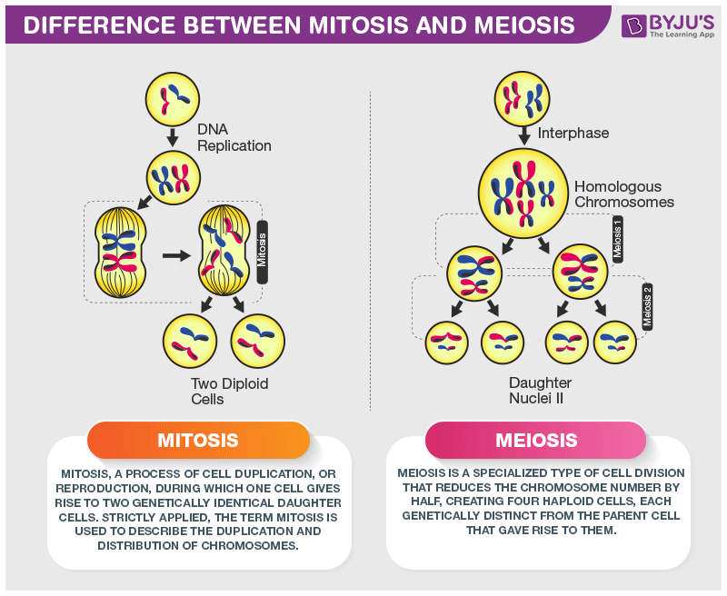
Image source: BYJU’S – Difference Between Mitosis and Meiosis (educational resource). Used for illustrative/educational purposes. Verify licensing and attribute from the original source.
Common Mistake: Students confuse the chromosome number outcome and the purpose of each division.
Correct Concept: Mitosis produces two genetically identical diploid daughter cells for growth and repair. Meiosis produces four genetically different haploid gametes for sexual reproduction.
Why Students Get Confused: Both processes involve similar-looking stages (prophase, metaphase, etc.) but differ in key events like crossing over and reduction division.
Tricky MDCAT MCQ: A diagram compares two cell division processes. One process shows one round of division producing two identical diploid cells. The other shows two rounds of division with crossing over and production of four haploid cells. Which statement correctly identifies both processes and their biological roles?
A. First process = meiosis (gamete formation); second process = mitosis (growth and repair). B. First process = mitosis (growth and repair, identical diploid cells); second process = meiosis (gamete formation, haploid cells with genetic variation). C. Both processes produce identical diploid cells. D. Both processes reduce chromosome number by half.
Answer: B
Explanation: Mitosis maintains chromosome number and produces identical cells for growth/repair. Meiosis halves the chromosome number and introduces variation through crossing over and independent assortment.
Quick Exam Tip: Mitosis = identical diploid (growth/repair). Meiosis = haploid + variation (gametes).
Diagram 22: Neuron structure
Image source: OpenStax Anatomy and Physiology – Nervous Tissue (educational open textbook). Used for illustrative/educational purposes. Verify licensing and attribute from the original source.
Common Mistake: Students mix up the direction of impulse travel or the function of dendrites versus axon.
Correct Concept: Dendrites receive impulses and carry them toward the cell body. The axon carries impulses away from the cell body to other neurons or effectors. Myelin sheath speeds up conduction.
Why Students Get Confused: The long axon and branched dendrites look similar in simplified diagrams.
Tricky MDCAT MCQ: A neuron diagram shows branched structures receiving signals and a long single process carrying signals away from the cell body, covered in a myelin sheath in places. Which statement correctly describes the direction of impulse and the role of the long process?
A. Branched structures (dendrites) receive impulses and carry them toward the cell body; the long process (axon) carries impulses away from the cell body. B. Branched structures carry impulses away from the cell body. C. The long process receives impulses. D. Myelin sheath slows down impulse conduction.
Answer: A
Explanation: Dendrites bring impulses to the cell body. The axon takes impulses away from the cell body. Myelin increases speed of conduction.
Quick Exam Tip: Dendrites = receive (toward cell body). Axon = send (away from cell body). Myelin = faster conduction.
Diagram 23: Synapse

Image source: Educational neuroscience resource – synaptic transmission diagram. Used for illustrative/educational purposes. Verify licensing and attribute from the original source.
Common Mistake: Students think the impulse crosses the synaptic cleft electrically or that neurotransmitters move in both directions.
Correct Concept: The impulse arrives at the presynaptic terminal, causes Ca²⁺ influx, vesicles release neurotransmitter into the synaptic cleft, and the neurotransmitter binds to receptors on the postsynaptic membrane, generating a new impulse.
Why Students Get Confused: The tiny gap and chemical nature of transmission are not obvious without clear arrows and labels.
Tricky MDCAT MCQ: A synapse diagram shows an impulse arriving at the end of one neuron, vesicles releasing a chemical into a narrow gap, and that chemical binding to receptors on the next neuron. Which statement correctly describes the sequence and the nature of transmission?
A. Electrical impulse jumps directly across the gap. B. Chemical neurotransmitter is released from presynaptic neuron into the synaptic cleft and binds to receptors on the postsynaptic neuron (chemical transmission). C. Neurotransmitter moves from postsynaptic to presynaptic neuron. D. The gap is bridged by direct cytoplasmic connection.
Answer: B
Explanation: Synaptic transmission is chemical. Neurotransmitter is released from the presynaptic side and acts on the postsynaptic side.
Quick Exam Tip: Synapse = chemical transmission via neurotransmitter. One-way (pre → post).
Diagram 24: Reflex arc

Image source: Educational biology resource – reflex arc diagram. Used for illustrative/educational purposes. Verify licensing and attribute from the original source.
Common Mistake: Students include the brain in every reflex or reverse the order of sensory and motor neurons.
Correct Concept: A reflex arc is a rapid, automatic response that bypasses the brain for speed: receptor → sensory neuron → relay neuron (in spinal cord) → motor neuron → effector.
Why Students Get Confused: Many diagrams show the spinal cord and brain together; students assume conscious involvement.
Tricky MDCAT MCQ: A diagram of a reflex shows a painful stimulus detected by receptors, impulse travelling via a sensory neuron to the spinal cord, a relay neuron connecting to a motor neuron, and the effector muscle contracting to withdraw the hand — all without involving the brain. Which statement correctly identifies the type of response and the reason for its speed?
A. Conscious voluntary action involving the brain. B. Reflex action – rapid and automatic because it bypasses the brain and uses a short spinal pathway. C. The brain must process every impulse before the muscle contracts. D. Only sensory neurons are involved.
Answer: B
Explanation: Reflexes are involuntary, rapid and protective because the pathway is short and does not require brain processing for the immediate response.
Quick Exam Tip: Reflex = receptor → sensory → relay → motor → effector (spinal cord shortcut, brain not needed for speed).


.jpg)

+by+Blacklotus+Landscaping.jpg)


0 Comments