
Chapter 21 Biology Class 12 RTS Important Short& Long Solved
1) Describe cell cycle with phases.
The cell cycle consists of two main phases: Interphase and Mitotic (M) phase. Interphase includes:

- G1 phase: Growth and preparation for DNA replication.
- S phase: DNA replication.
- G2 phase: Further growth and preparation for mitosis.
- The M phase includes:
- Mitosis: Division of the nucleus (Prophase, Metaphase, Anaphase, Telophase).
- Cytokinesis: Division of the cytoplasm.
2) Why interphase is called resting phase?
Interphase is called the "resting phase" because the cell does not undergo visible division during this phase, although it is actively growing and performing functions. The term is misleading as the cell is preparing for division.
3) What is mitotic apparatus?
The mitotic apparatus consists of the structures responsible for chromosome movement during mitosis, primarily the spindle fibers, centrioles, and asters.
4) How cytokinesis take place in plants?
In plant cells, cytokinesis occurs through the formation of a cell plate, which develops at the center of the cell and eventually forms a new cell wall, separating the two daughter cells.
5) Define metastasis.
Metastasis is the spread of cancer cells from their original (primary) site to other parts of the body, forming secondary tumors.
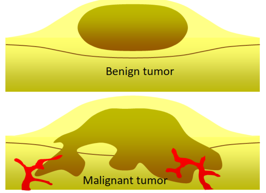
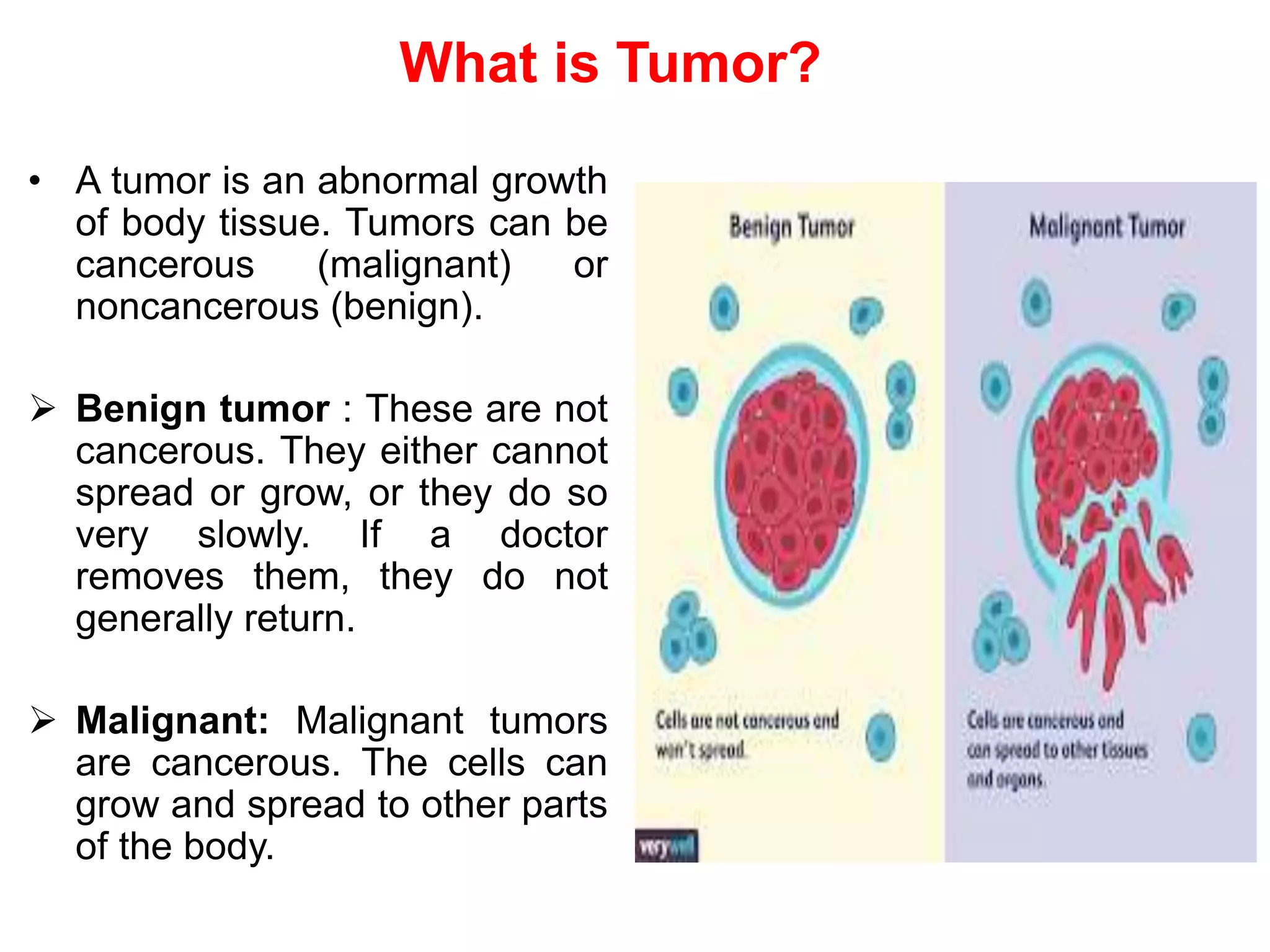
6) Difference between Benign and malignant tumor?
- Benign tumors: Non-cancerous, localized, do not spread.
- Malignant tumors: Cancerous, can invade nearby tissues and spread to other parts of the body (metastasis).
7) Enlist stages of prophase 1 of meiosis.
The stages of Prophase 1 of meiosis are:
- Leptotene: Chromosomes begin to condense.
- Zygotene: Homologous chromosomes pair up (synapsis).
- Pachytene: Crossing over occurs.
- Diplotene: Chromosomes start to separate but remain attached at chiasmata.
- Diakinesis: Chromosomes are fully condensed, and nuclear membrane breaks down.
8) Give importance of meiosis.
Meiosis is essential for sexual reproduction as it produces haploid gametes (sperm and egg), ensuring genetic diversity and maintaining the chromosome number across generations.
9) Define nondisjunction with causes.
Nondisjunction is the failure of chromosomes to separate properly during cell division, leading to an abnormal number of chromosomes in the daughter cells. It can be caused by errors during anaphase of mitosis or meiosis.
10) Explain Down's syndrome.
Down's syndrome is a genetic disorder caused by an extra copy of chromosome 21 (trisomy 21), resulting in developmental and physical delays, characteristic facial features, and possible intellectual disabilities.
11) Difference between apoptosis and necrosis.
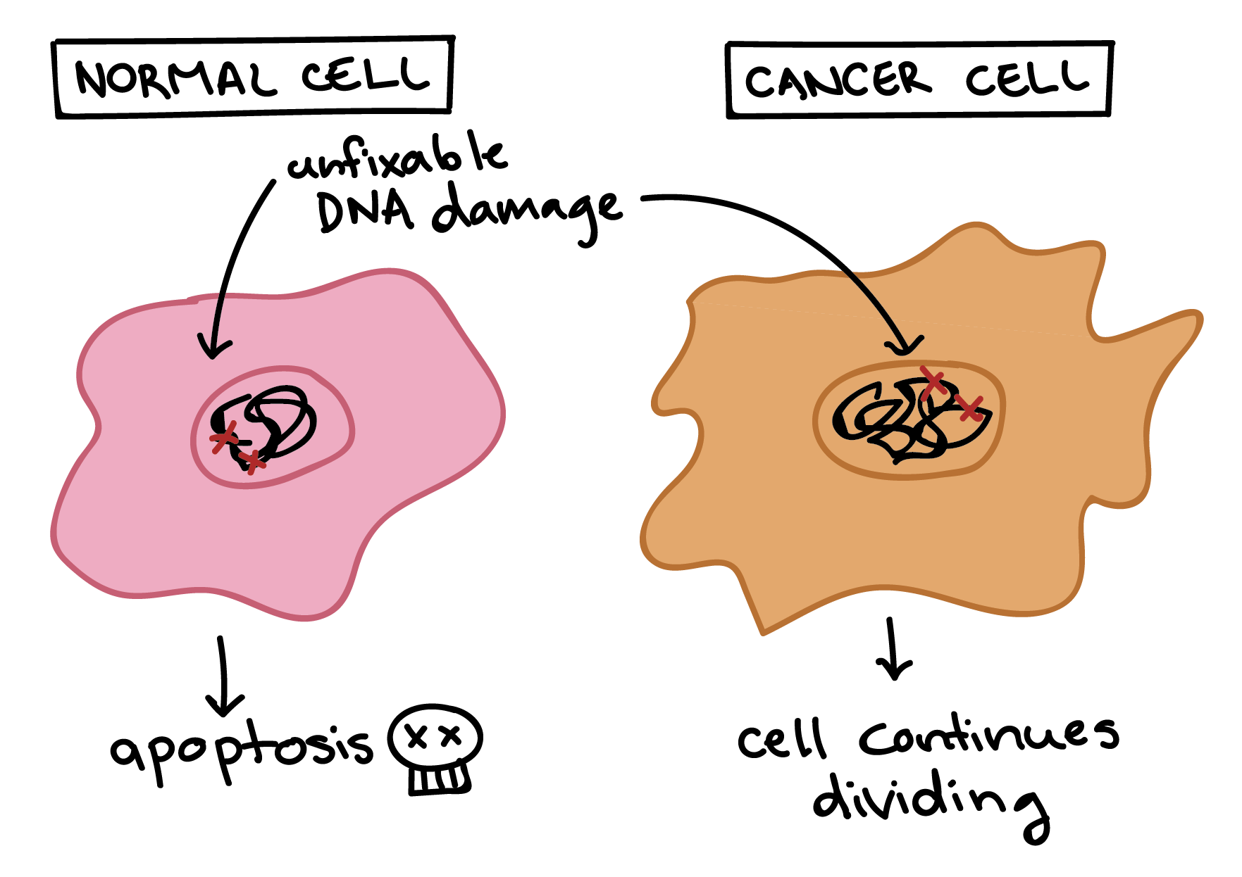
- Apoptosis: Programmed cell death, a controlled process that occurs for normal development or response to damage.
- Necrosis: Uncontrolled cell death due to injury, often leading to inflammation and tissue damage.
Write a note on cancer.
Definition Cancer is an abnormal and uncontrolled multiplication (proliferation) of cells that results in the formation of a mass of cells called a tumour. In normal cells, growth is highly regulated and coordinated. Normal cells exhibit contact inhibition — when they come into contact with other cells, they stop dividing. Cancer cells lose this property, leading to uncontrolled growth and tumour formation.
:max_bytes(150000):strip_icc()/illo_normal-cells-cancer-cells-596cdd256f53ba00111a65bb.png)
Types of Tumours Tumours are of two main types:
- Benign Tumours
- Confined to their original location.
- Do not spread to other parts of the body.
- Grow slowly.
- Cause little damage.
- Usually non-cancerous and can be removed by surgery; they do not regrow easily.
- Malignant Tumours (True Cancer)
- Grow rapidly.
- Invade and damage surrounding normal tissues.
- Compete with normal cells for nutrients, starving them.
- Cells from the tumour can slough off, enter blood or lymph, and form new tumours at distant sites. This property is called metastasis — the most feared characteristic of cancer.
Causes of Cancer (Carcinogens) Cancer is caused by physical, chemical, or biological agents called carcinogens. These transform normal cells into cancerous cells by activating proto-oncogenes (c-onc or cellular oncogenes) into oncogenes.
- Physical agents: Ionising radiations (X-rays, gamma rays) and non-ionising radiations (UV rays) — cause DNA damage.
- Chemical agents: Tobacco smoke (main cause of lung cancer), nicotine, caffeine, vinyl chloride, mustard gas, etc.
- Biological agents: Oncogenic viruses (contain viral oncogenes), certain parasites.
Differences between Normal Cells and Cancer Cells
| Characteristic | Normal Cells | Cancer Cells |
|---|---|---|
| Growth & Division | Controlled; stop at contact inhibition; undergo apoptosis if damaged | Uncontrolled; no contact inhibition; no apoptosis |
| Shape & Size | Uniform | Irregular shape & size, large nucleus, less cytoplasm |
| Communication | Communicate properly | Poor or no communication |
| Adhesion & Spread | Stick to other cells; do not spread | Lose adhesion; metastasise via blood/lymph |
| Differentiation | Mature and specialised | Remain immature |
Cancer Detection and Diagnosis (Very Important for Exams) Early detection increases chances of successful treatment.
- Biopsy: A small piece of suspected tissue is taken, stained, and examined under a microscope (histopathological studies).
- Blood & Bone Marrow Tests: For leukaemia (increased WBC count).
- Imaging Techniques:
- Radiography (X-rays)
- CT (Computed Tomography) scan — 3D image using X-rays
- MRI (Magnetic Resonance Imaging) — uses strong magnetic fields & non-ionising radiations
- Antibodies against cancer-specific antigens.
- Molecular biology techniques to detect cancer-related genes.
Treatment of Cancer Most cancers are treated by a combination of methods:
- Surgery: Removal of the tumour (effective for localised/benign tumours).
- Radiotherapy: Tumour cells are irradiated lethally (high-energy radiation) without much damage to surrounding normal cells.
- Chemotherapy: Use of anti-cancer drugs/chemicals that kill rapidly dividing cancer cells (side effects: hair loss, anaemia, nausea).
- Immunotherapy: Biological response modifiers (e.g., α-interferon) are given to activate the patient's immune system to destroy tumour cells.
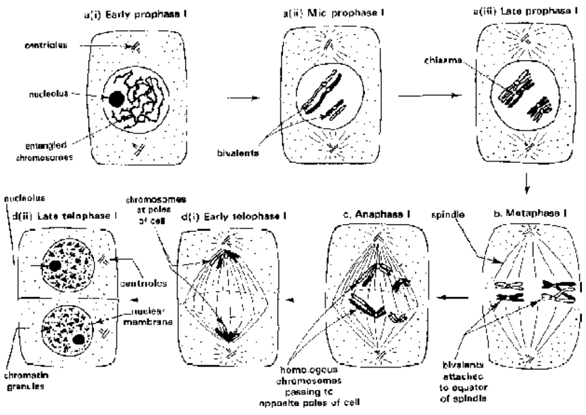
Describe prophase 1 of meiosis in details
Prophase I of Meiosis Detailed Long Note for Class 12 Biology (Important Exam Question – RTS/HED/Punjab Board/NCERT Pattern)
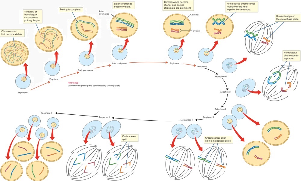
Prophase I is the longest and most complex phase of meiosis I. It is divided into five sequential substages: Leptotene, Zygotene, Pachytene, Diplotene, and Diakinesis. During this phase, homologous chromosomes pair, synapsis occurs, and crossing over takes place, which is responsible for genetic recombination. This phase is unique to meiosis and does not occur in mitosis.
Below is the detailed description of each substage with important chromosomal events:
1. Leptotene (Leptonema)
- Chromosomes become gradually visible as long, thin, slender threads (hence called leptotene = thin thread).
- Chromosomes start condensing but are still elongated and intertwined.
- Each chromosome has already replicated in the preceding S-phase, so it consists of two sister chromatids joined at the centromere (but chromatids are not yet clearly visible).
- Nuclear membrane and nucleolus are still intact.
- Centrioles (in animal cells) begin to move towards opposite poles.


2. Zygotene (Zygonema)
- Homologous chromosomes start pairing (synapsis) with each other.
- Pairing begins at one or more points and proceeds in a zipper-like manner.
- The paired homologous chromosomes are called bivalents (or tetrads because each bivalent has 4 chromatids).
- A synaptonemal complex (proteinaceous structure) forms between the paired homologues, holding them tightly together.
- Nuclear membrane and nucleolus are still present.


3. Pachytene (Pachynema)
- This is the most important substage for exams.
- Bivalents become short and thick.
- Synapsis is complete; each bivalent now clearly shows four chromatids (tetrad stage).
- Crossing over (exchange of genetic material) occurs between non-sister chromatids of homologous chromosomes at several points.
- The points of crossing over appear as X-shaped structures called chiasmata (singular: chiasma).
- This crossing over leads to genetic recombination, which is the main source of variation in sexually reproducing organisms.
- Synaptonemal complex is fully formed.
- Nucleolus and nuclear membrane still intact.
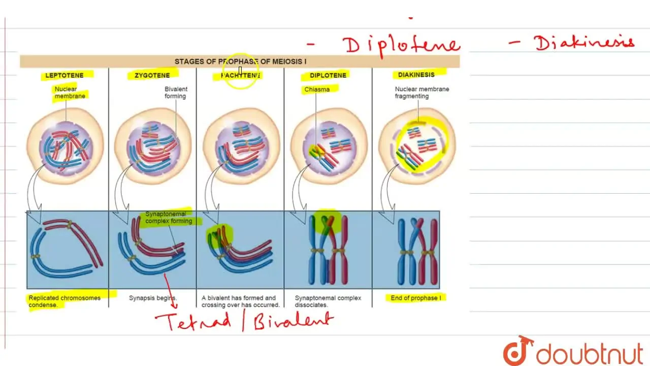
4. Diplotene (Diplonema)
- Synaptonemal complex begins to dissolve and disappears.
- Homologous chromosomes start separating (repulsion) except at the points of chiasmata.
- Chiasmata become clearly visible and start terminalising (moving towards the ends of chromosomes).
- Chromosomes appear as bivalents with 4 chromatids held together only at chiasmata.
- In many organisms (especially females of vertebrates), this stage is very long and may last for months or years (e.g., oocytes remain in diplotene till ovulation).

5. Diakinesis (Diakinesis)
- Final stage of prophase I.
- Chromosomes become highly condensed, short, and thick.
- Chiasmata terminalise completely (move to the tips of chromosomes).
- Nucleolus disappears completely.
- Nuclear membrane breaks down (fragmentation starts).
- Spindle fibres begin to appear.
- Bivalents (tetrads) are now ready to move towards the metaphase plate.
- This marks the end of prophase I.
Summary Table (Very Important for Exams – 5–8 Marks Question)
| Substage | Key Events | Visible Structures | Nuclear Membrane & Nucleolus |
|---|---|---|---|
| Leptotene | Condensation begins | Thin threads, no pairing | Present |
| Zygotene | Synapsis (pairing) of homologues | Bivalents, Synaptonemal complex | Present |
| Pachytene | Crossing over, genetic recombination | Tetrads, Chiasmata appear | Present |
| Diplotene | Synaptonemal complex dissolves, repulsion | Chiasmata visible | Present |
| Diakinesis | Maximum condensation, terminalisation | Thick bivalents, terminal chiasmata | Disappear |
Important Points to Remember for Long Questions
- Prophase I is divided into 5 substages — always write names in order: Leptotene → Zygotene → Pachytene → Diplotene → Diakinesis.
- Pachytene is the most significant substage because crossing over occurs here.
- Chiasmata are the morphological evidence of crossing over.
- Synaptonemal complex forms in Zygotene and dissolves in Diplotene.
- This phase is responsible for reduction in chromosome number (from 2n to n in meiosis I) and genetic variation.


.jpg)


+by+Blacklotus+Landscaping.jpg)


0 Comments