
Funaria hygrometrica Detailed Life Cycle
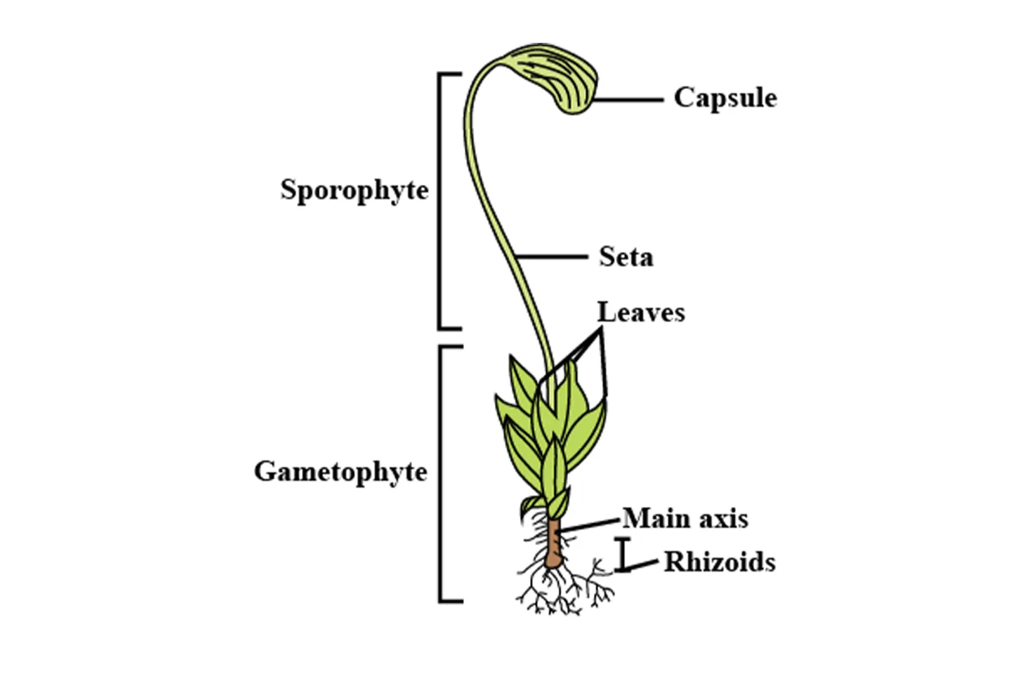
1. Bryophyta - Funaria Gametophyte
The gametophyte is the haploid (n) phase, dominant and photosynthetic. In Funaria, it's an acrocarpous (upright, unbranched) type of leafy gametophyte that develops from the protonema(Protonema is a thread-like, green, haploid juvenile stage in the life cycle of mosses (Bryophytes) that develops from a germinating spore and later gives rise to the leafy gametophyte.


It is poikilohydric (desiccation-tolerant), with adaptations for disturbed habitats.
Development of Gametophyte
Spore Germination (Spore → Protonema)
Spore germination marks the first stage of the gametophytic generation in mosses. The haploid spore, measuring 12–20 μm in diameter, possesses a thick exine often ornamented with papillae, which provides protection during dormancy.
Under favorable environmental conditions—specifically adequate moisture, a temperature range of 20–25°C, and high humidity—the spore resumes metabolic activity and germinates.
Germination Process
- The spore wall ruptures, and a germ tube consisting of 1–2 cells emerges.
- This germ tube elongates and differentiates into chloronema, which is composed of green, branched, filamentous cells rich in chloroplasts.
- Chloronema represents the photosynthetic phase of the developing protonema.
Differentiation of Rhizoids
- From the protonemal filaments, rhizoidal branches arise.
- These branches are brown in color, non-photosynthetic, and function in anchorage and absorption.
- The cells of rhizoidal filaments show oblique septa, a distinguishing feature from chloronema cells.
Cellular Organization
- Growth of the protonema is controlled by a prominent apical cell.
- The apical cell is triangular in shape and divides regularly to add new segments, resulting in filament elongation and branching.
Time Duration
- Complete formation of a well-developed protonema occurs within 3–7 days after spore germination, depending on environmental conditions.
Vegetative Propagation
- The protonema may produce gemmae, which are multicellular buds (10–30 cells).
- These gemmae detach and develop into new protonemata, enabling asexual (vegetative) reproduction.
Evolutionary Significance
-
The protonema stage is considered homologous to filamentous green algae, reflecting the evolutionary origin of bryophytes from algal ancestors.

Stage 2: Bud Formation & Gametophore Initiation
(Protonema → Leafy Shoot)
After the establishment of a well-developed protonema, the next phase in the moss life cycle is bud formation, which leads to the development of the leafy gametophyte (gametophore).
Bud Formation
- Side branches of the protonema give rise to small, multicellular buds.
- Each bud initially consists of 3–4 cells and represents the gametophore initial.
- These buds undergo a characteristic octant stage, similar to early embryonic development in higher plants.
Octant Stage & Patterning
- During the octant stage, the bud divides to form eight cells arranged in a precise pattern.
- This stage establishes apical–basal polarity:
- Epibasal region → develops into the leafy shoot
- Hypobasal region → forms rhizoids
- This organization is crucial for correct gametophyte architecture.
Development of Gametophore
-
The bud differentiates into:
-
A central stem axis
- A rosette of rhizoids at the base
- Leaves arise on the stem in a spiral phyllotaxy, a typical bryophytic feature.
- Complete transformation from protonema to a young gametophore takes 2–4 weeks, depending on environmental conditions.
Hormonal Control
- Auxin gradients play a central role in:
- Establishing cell polarity
- Directing apical growth
- Regulating bud differentiation into shoot and rhizoid regions
Ultrastructural Features
- Cells of the developing gametophore possess well-developed chloroplasts.
- Chloroplasts show stacked grana, indicating active C₃ photosynthesis.
- This marks the transition from a filamentous to a fully photosynthetic leafy phase.
Pro Tip: Culture on Knop's agar for protonema induction; track development under stereomicroscope.

2. Bryophyta - Funaria - Male Head (Antheridial Head)
The male head is the perigonium – a cluster of antheridia on the apical main axis. Protandrous (antheridia mature first).

-
Development begins from an apical gametophyte cell, which differentiates into a superficial initial of dermal origin.
This initial divides periclinally to form:
- An outer jacket layer (single-cell thick)
- Inner androgonial mother cells
- The jacket layer provides mechanical protection, while the inner cells remain meristematic.
- This stage occurs 1–2 weeks after gametophore emergence.
- Site of development: The male head is terminal on the main axis of the leafy gametophyte.
-
Androgonial mother cells divide repeatedly by mitosis (not meiosis).
A compact androgonial mass of about 100–200 cells is formed.
-
Each androgonial cell ultimately differentiates into a single antherozoid.
-
Antherozoids are:
-
Biflagellate
10–15 μm long
Spirally coiled
- Provided with two anterior flagella
-
They are haploid and motile, adapted for movement in a water film.
- Jacket cells elongate during maturation.
- Chromoplasts develop in jacket cells, contributing to protection and structural support.
- Paraphyses, which are 4–6 celled and capitate, are present among the antheridia.
Development of Male Head (Antheridial Head)
The male reproductive structure in mosses develops at the apex of the gametophyte and bears numerous antheridia, collectively forming the male head (antheridial head).
Stage 1: Initial Differentiation
Stage 2: Androgonial Division & Antherozoid Formation
Androgonial Divisions
Antherozoids
Jacket Layer & Paraphyses
Paraphyses function in:
- Nutrition
- Secretion of mucilage
- Protection against desiccation
- Antherozoids possess a well-developed flagellar apparatus with a typical 9+2 microtubule arrangement.
- Each antheridium opens by an operculum-like pore.
- Dehiscence occurs due to mucilage swelling, allowing the release of antherozoids in water.
- Moss antherozoids represent a reduced condition compared to multiflagellate ancestors seen in some lower plants.
- Fertilization is aided by chemotaxis, where antherozoids are attracted towards the archegonium by sucrose gradients.
- This reflects an evolutionary adaptation for efficient fertilization in terrestrial habitats.
Ultrastructural Features
Evolutionary Significance

3. Bryophyta - Funaria - Female Head and Fertilization (Archegonial Head & Fertilization)
🌿 Development of Female Head (Archegonial Head)
Stage 1: Archegonial Initial & Differentiation
-
A superficial cell on a short lateral branch of the gametophyte differentiates into an archegonial initial.
-
The archegonial initial divides to form:
-
Neck canal cells (usually 6–10, elongated and slightly twisted)
Venter, containing:
Egg cell
- Ventral canal cell
The archegonium develops a double jacket layer:
- Outer jacket
- Inner jacket
- Time: Appears about 1 week after antheridia formation
- Site: On elongated lateral branches arising from the base of the gametophyte
Stage 2: Maturation of Archegonium
Neck canal cells degenerate, forming a mucilage-filled canal
This mucilage acts as a chemotactic attractant for antherozoids
- Ventral canal cell disintegrates
- Egg nucleus enlarges and becomes metabolically active
- The mature archegonium becomes flask-shaped, about 300 µm long
Archegonia are:
- Embedded within perichaetial bracts
- Surrounded by paraphyses, providing protection and moisture retention
Ultrastructural Features of Egg Cell
- Large central vacuole
- Dense cytoplasm rich in organelles
- Presence of plasmodesmata, enabling symplastic nutrition from surrounding tissues
- Indicates matrotrophic dependence of the developing zygote
🌧️ Fertilization Process
Mechanism
- Fertilization requires a thin film of water (rain or splash dispersal)
- Biflagellate antherozoids swim toward the archegonium
- Sucrose-based chemotaxis from archegonial mucilage guides sperm entry
- Only one antherozoid penetrates the archegonium neck
- Male and female nuclei fuse, forming a diploid zygote inside the venter
Time Frame
Fertilization occurs within hours of antherozoid release
Protective & Advanced Features
-
After sperm entry, a mucilage plug forms in the neck, preventing further entry
Polyspermy is blocked by:
Membrane depolarization of the egg
- The zygote remains retained within the venter
- Nutrients are supplied by the gametophyte → matrotrophic nutrition
4. Bryophyta - Funaria - Sporophyte
Delopment of Sporophyte in Funaria
Stage 1: Embryogeny (Zygote → Embryo)
Overview: Embryogeny is the initial phase of sporophyte development, starting right after fertilization in the archegonium of the female gametophyte. In Funaria, the zygote (diploid, 2n) forms from the fusion of sperm (n) and egg (n). This stage transforms the zygote into a multicellular embryo over about 1 week. It's a critical period of cell division and differentiation, establishing the basic body plan: the sporophyte will be dependent on the gametophyte for nutrition via haustorial cells.
Detailed Process:
- Zygote Formation: The zygote is embedded in the venter of the archegonium. It's a single diploid cell with a large nucleus and dense cytoplasm. No immediate division occurs; it absorbs nutrients from the surrounding gametophyte tissue.
- First Division: The zygote undergoes its first mitotic division, which is transverse (perpendicular to the archegonial axis) in Funaria (unlike vertical in some ferns). This produces a 2-celled stage:
- Epibasal cell (upper, terminal): Smaller, cytoplasm-rich; precursor to the capsule (spore-bearing part).
- Hypobasal cell (lower, basal): Larger; gives rise to the foot and seta (stalk).
- Octant Stage: The epibasal cell divides transversely to form a quadrant (4 cells), then each divides anticlinally to reach the octant stage (8 cells). The hypobasal cell divides to form a tier. This sets up polarity:
- Epibasal region (upper octant): Develops into capsule precursors (future theca and apophysis).
- Hypobasal region (lower tier): Forms foot and seta precursors.
- Young Embryo Formation: Further periclinal and anticlinal divisions create a globular embryo. Suspensor-like cells (from hypobasal derivatives) elongate and push the embryo upward, aiding nutrient transfer from the gametophyte (like a primitive placenta). These cells degenerate later.
- Timeline: Completes in ~1 week under optimal moist conditions.
- Genetic Aspects: Biparental gene expression dominates; however, imprinting occurs—paternal genes promote growth (e.g., cell expansion), while maternal genes control early patterning. This ensures balanced diploid development.
- Key Functions: Establishes apical-basal axis; suspensor facilitates nutrient uptake via plasmodesmata connections.
- Evolutionary Note: In bryophytes like Funaria, embryogeny is "reduced" compared to vascular plants—no true suspensor like in angiosperms, but analogous cells ensure embryo nutrition.
Labelled Diagram: Sequential diagram showing embryogeny stages in Funaria (L.S. view). Note the transverse first division and regional differentiation.

(Reference: From Uttarakhand Open University botany notes—clear labels for zygote, 2-celled, octant, and young embryo with epibasal/hypobasal regions and suspensor.)
Stage 2: Organ Differentiation (Embryo → Foot-Seta-Capsule)
Overview: This phase follows embryogeny (~3–6 weeks) and involves organogenesis, where the young embryo differentiates into the tripartite sporophyte: foot (anchoring/absorptive), seta (elongating stalk), and capsule (spore-producing). The entire sporophyte remains attached to and nourished by the female gametophyte. Differentiation is driven by meristems and hormones like auxin.
Detailed Process:
- Foot Development: The basal hypobasal region expands into the foot (haustorial in nature). It's embedded in the archegonial venter tissue. Ultrastructure includes transfer cells with wall ingrowths (labyrinthine) for efficient solute uptake (e.g., sugars, amino acids) from the gametophyte via symplastic pathways. The foot has rhizoid-like projections for anchorage.
- Seta Formation: From the middle embryonic tier, the seta arises via an intercalary meristem (2–3 cell layers). Auxin gradients drive elongation and twisting (hygroscopic, due to asymmetric cell wall thickening—helps in spore dispersal later). Ultrastructure: Central leptoids (conducting cells) degenerate before meiosis, while hydroids (water-conducting) persist in the columella.
- Capsule Differentiation: The epibasal region forms the capsule, divided into:
- Apophysis (basal swollen part): Photosynthetic with stomata for gas exchange and transpiration; contributes ~30% of carbon needs via photosynthesis.
- Theca (main spore sac): Houses diploid sporogenous tissue that undergoes meiosis to form haploid spores.
- Operculum (lid): Caps the theca; detaches during dehiscence.
- Calyptra Role: Derived from the archegonial neck (epigonium), it's a hairy, protective covering over the young capsule. It enlarges with growth and ruptures later, preventing desiccation.
- Timeline: 3–6 weeks; seta elongates rapidly in moist conditions.
- Ultrastructure Highlights: Seta has hydroid-filled columella (sterile central axis); capsule wall has amphithecium-derived layers. Peristome (arthrodontous type) develops from peristomial membrane—exostome teeth are hygroscopic (bend with moisture changes via cellulose microfibrils).
- Evolutionary Note: Bryophyte sporophytes are "reduced" vs. vascular plants (no leaves/roots), but the twisted seta and peristome are adaptations for wind dispersal in terrestrial habitats. Funaria's arthrodontous peristome is primitive, aiding controlled spore release.
Labelled Diagram: L.S. of mature sporophyte in Funaria, highlighting organ parts and key structures.

(Reference: From VPScience Funaria life cycle PDF—detailed L.S. with foot, twisted seta, capsule components, haustorium, hydroids, stomata, and peristome teeth.)
Stage 3: Maturation & Dehiscence
Overview: The final phase focuses on spore maturation inside the capsule and mechanisms for dehiscence (spore release). This ensures dispersal of 10⁴–10⁵ haploid spores per capsule, completing the alternation of generations. Maturation takes additional weeks post-differentiation, triggered by environmental cues like drying.
Detailed Process:
- Maturation:
- Inside the theca, sporogenous cells (diploid) undergo meiosis → tetrads → haploid spores (with exine for protection).
- Calyptra remains intact initially, protecting against desiccation. Apophyseal stomata open for transpiration and CO₂ uptake, supporting photosynthesis (up to 30% of sporophyte's C fixation).
- Peristome teeth mature: Exostome (outer) teeth are hygroscopic, bending outward in dry air due to microfibril orientation.
- Dehiscence Mechanism:
- Calyptra Rupture: As the capsule matures, the calyptra splits and is shed (or pushed up by elongating seta), exposing the operculum.
- Dry Conditions Trigger: Seta untwists (reverses hygrocastic movement), elevating the capsule for better dispersal.
- Peristome Opening: Teeth hygroscopically bend (via annular thickenings), creating gaps for spores to escape. Operculum detaches via a separation layer.
- Spores (10⁴–10⁵ per capsule, ~20–30 μm diameter) are released in bursts, aided by air currents. Annulus (ring below operculum) prevents premature release.
- Timeline: Peaks 4–8 weeks after fertilization; full dehiscence in dry weather.
- Functions: Ensures spore viability and wide dispersal; apophyseal photosynthesis reduces gametophyte dependency.
- Evolutionary Note: Hygroscopic mechanisms (seta twist, peristome) are bryophyte innovations for xeric adaptation, unlike simple capsules in algae. In Funaria, this supports colonization of disturbed soils (e.g., burnt areas).
Labelled Diagram: Diagram illustrating maturation, calyptra rupture, and peristome-mediated dehiscence in Funaria sporophyte.
(Reference: From PW Live NEET notes on Funaria—shows capsule maturation, calyptra, operculum detachment, and spore release with peristome details.)
5. Bryophyta - Funaria - Alternation of Generations
Haplo-diplontic life cycle: Isomorphic but gametophyte dominant (independent, photosynthetic) vs. sporophyte dependent (matrotrophic, elevated for dispersal).
Developmental Sequence & Alternation
| Generation | Ploidy | Duration | Key Developments | Transition |
|---|---|---|---|---|
| Gametophyte | n | 1–3 months (ephemeral) | Spore → protonema → buds → leafy shoot → sex organs. Vegetative: Gemmae/apospory. | Fertilization (n + n → 2n zygote). |
| Sporophyte | 2n | 1–2 months | Zygote → embryo (octant) → foot-seta-capsule → meiosis in theca → spores. | Meiosis → n spores → new gametophyte. |
| Alternation | n ↔ 2n | Cycle: 2–3 months | Dependent sporophyte on gametophyte; semi-autonomy via stomata/seta. | Water-dependent; genetic conflicts (paternal bias for longer seta). |
- Full Cycle: Spore germination → gametophyte → gametogenesis → fertilization → embryogeny → sporogenesis (meiosis) → spore dispersal → repeat.
Evolutionary Insight: Bryophyte model for land plant transition; sporophyte elaboration (seta elongation) key innovation.
Advanced: Genomic imprinting resolves parent-offspring conflict; monoecy favors selfing (shorter cycles in disturbed sites).


.jpg)


0 Comments