
Short and Long Answers on Reproduction and Growth

-
Give two disadvantages of cloning:
- Cloning reduces genetic diversity, making populations more vulnerable to diseases.
- Clones may be more susceptible to environmental changes or health issues due to a lack of genetic variation.
-
Difference between sexual and asexual reproduction:
- Sexual reproduction involves the fusion of male and female gametes, resulting in genetically diverse offspring.
- Asexual reproduction involves a single organism reproducing without the involvement of gametes, producing genetically identical offspring.
-
How evolution of pollen tube is important for spermatophytes?
-
The pollen tube allows the sperm to travel to the ovule for fertilization, which is critical for the reproduction of seed-bearing plants (spermatophytes).
-
-
Define parthenocarpy with example:
-
Parthenocarpy is the formation of fruit without fertilization. Example: Seedless bananas.
-
-
Define fruit set:
-
Fruit set is the process during which the fertilized ovules develop into fruit.
-
-
Define periodism:
-
Periodism refers to the synchronization of reproductive cycles in plants with environmental factors like light and temperature, influencing flowering and fruiting.
-
-
Define vernalization with its importance in plants:
-
Vernalization is the exposure of plants to cold temperatures to induce flowering, especially in winter-hardy species. It is important for ensuring proper timing of reproduction.
-
-
Difference between short day and long day plants:
-
Short day plants flower when the day length is shorter than a critical period.
-
Long day plants flower when the day length exceeds a certain threshold.
-
-
Define Apomixis:
-
Apomixis is a form of asexual reproduction in plants where seeds are produced without fertilization, resulting in offspring genetically identical to the parent.
-
-
Difference between diploid parthenogenesis and haploid parthenogenesis:
- Diploid parthenogenesis produces offspring with the same chromosome number as the parent (diploid).
- Haploid parthenogenesis produces offspring with half the chromosome number (haploid).
Additional Questions (from continuation of Ch#18 Reproduction)
-
Difference between identical and fraternal twins:
- Identical twins are formed from a single fertilized egg that splits, resulting in genetically identical individuals.
- Fraternal twins result from two separate eggs fertilized by two separate sperm, leading to genetically unique siblings.
-
Difference between oviparous and viviparous:
-
Oviparous animals lay eggs that hatch outside the body.
-
Viviparous animals give birth to live young that develop inside the mother.
-
-
Difference between external and internal fertilization:
-
External fertilization occurs outside the female's body, as seen in fish and amphibians.
-
Internal fertilization occurs inside the female’s body, typical of mammals, reptiles, and birds.
-
-
What is ovoviviparity?
-
Ovoviviparity is a reproductive strategy where eggs develop inside the mother’s body but hatch before being laid, as seen in some fish and reptiles.
-
-
What is the function of Sertoli cells?
-
Sertoli cells support and nourish developing sperm cells in the testes.
-
-
Define ovulation:
-
Ovulation is the release of an egg from the ovary, which can be fertilized by sperm.
-
-
What is menopause? At what age does it start?
-
Menopause is the cessation of a woman's menstrual cycle, typically occurring between the ages of 45 and 55.
-
-
What is after birth?
-
After birth refers to the period following childbirth, including processes like the delivery of the placenta and recovery.
-
-
What is test tube babies?
-
Test tube babies are children born through in vitro fertilization (IVF), where eggs and sperm are combined outside the body and then implanted in the mother's womb.
-
-
Write a note on the female reproductive system:
-
The female reproductive system includes the ovaries, fallopian tubes, uterus, and vagina, responsible for egg production, fertilization, and childbirth.
-
-
Write a note on the menstrual cycle:
-
The menstrual cycle is a monthly process where the body prepares for pregnancy. It involves hormonal changes leading to ovulation, followed by menstruation if fertilization does not occur.
-
-
Discuss the process of birth:
-
The process of birth (parturition) involves labor, during which the cervix dilates, the baby moves through the birth canal, and the placenta is delivered after the baby.
-
-
Write a note on STD:
-
Sexually transmitted diseases (STDs) are infections passed from one person to another through sexual contact. Common STDs include chlamydia, gonorrhea, and HIV.
-
Ch#19 Growth and Development
-
Define gray crescent with importance:
-
The gray crescent is a region in the fertilized egg of some species where the future dorsal side of the embryo develops. It plays a critical role in the formation of the embryo's body axes.
-
-
Define embryonic induction:
-
Embryonic induction is the process by which certain cells influence the development of nearby cells, directing the formation of specific tissues and organs.
-
-
Define aging with symptoms:
-
Aging refers to the gradual decline of bodily functions, often marked by wrinkles, decreased mobility, and reduced organ function.
-
-
How aging can be slowed down?
-
Aging can be slowed down through a healthy diet, regular exercise, managing stress, and avoiding environmental toxins.
-
-
Difference between gerontology and teratology:
-
Gerontology is the study of aging and its effects on the body.
-
Teratology is the study of abnormal development, particularly birth defects.
-
-
What are neoblasts?
-
Neoblasts are undifferentiated cells capable of regenerating lost body parts, found in certain invertebrates like planarians.
-
-
What are teratogens? Give examples:
-
Teratogens are substances that cause birth defects, such as alcohol, drugs, and certain infections.
-
-
Write a note on aging:
-
Aging is a natural biological process characterized by the gradual decline in cellular function, leading to reduced physical and mental capabilities.
-
-
What is regeneration, explain with different examples:
-
Regeneration is the process by which organisms can regrow lost or damaged body parts, such as starfish regenerating limbs or humans growing new skin after injury.
-
-
Discuss various types of abnormalities in development:
-
Abnormalities in development can include congenital disorders, such as cleft lip, Down syndrome, and neural tube defects, caused by genetic mutations, environmental factors, or infections
-
Long Questions with answers
FRUIT SET
Comprehensive Guide to Chapter 18 & 19 RTS Biology Questions: Short and Long Answers on Reproduction and Development
Definition Fruit set is the process by which a flower, after successful pollination and fertilization, develops into a young fruit through the enlargement of the ovary.

Detailed Explanation (Step-by-Step)
- Pollination: Pollen grains are transferred from the anther to the stigma.
- Fertilization: The pollen tube grows through the style and enters the ovule, where double fertilization occurs. One male gamete fuses with the egg cell to form the zygote (embryo), while the second fuses with the central cell to form the triploid endosperm.
- Hormonal Stimulation: The developing seeds produce hormones such as auxin, gibberellins and cytokinins. These hormones promote rapid cell division and enlargement of the ovary wall (pericarp).
- Result: The ovule develops into a seed and the ovary develops into a fruit.
Parthenocarpy (Important for exams) Development of fruit without fertilization, resulting in seedless fruits (e.g., banana, seedless grapes). It can occur naturally or can be induced by spraying auxin or gibberellin hormones.
Key Points
- Fruit set prevents flower drop.
- Optimal temperature for good fruit set is 20–30 °C.
- Poor pollination or fertilization leads to low fruit set.

FRUIT RIPENING
Definition Fruit ripening is the final stage of fruit development in which the fruit becomes soft, sweet, coloured and aromatic to facilitate seed dispersal. It is primarily regulated by the plant hormone ethylene.
Types of Ripening
- Climacteric fruits (e.g., apple, banana, mango, tomato): Show a sudden climacteric rise in respiration rate and a burst of ethylene production.
- Non-climacteric fruits (e.g., citrus, strawberry, grapes): Ripen gradually without any ethylene burst or respiratory peak.
Biochemical Changes During Ripening
- Colour: Chlorophyll is broken down; carotenoids (yellow/orange) or anthocyanins (red/purple) are synthesised.
- Texture: Enzymes (polygalacturonase, pectin methylesterase) degrade pectin in the cell wall, causing softening.
- Taste: Starch is converted into simple sugars (sweetness increases); organic acids decrease.
- Aroma: New volatile compounds (esters, aldehydes) are produced.
Ethylene Biosynthesis (Important for long questions) Methionine → S-adenosyl methionine → ACC (by ACC synthase) → Ethylene (by ACC oxidase). Ethylene binds to receptors and activates ripening genes.
Commercial Applications
- Ethylene gas is used for artificial ripening (e.g., in banana ripening chambers).
- 1-MCP (1-methylcyclopropene) is sprayed to block ethylene receptors and delay ripening during storage and transport.

MALE HUMAN REPRODUCTIVE SYSTEM
Definition The male reproductive system is a group of organs that produce, store, nourish and deliver male gametes (sperms) into the female reproductive tract during copulation. It also secretes the male sex hormone testosterone.
Main Functions
- Spermatogenesis (sperm production)
- Secretion of testosterone
- Transfer of sperms to female body
Parts of the System & Their Functions (Teach point-wise with diagram)
- Testes (Primary sex organs)
- Pair of oval glands (4–5 cm) present in scrotum.
- Temperature 2–2.5°C lower than body temperature (essential for sperm formation).
- Seminiferous tubules → site of spermatogenesis.
- Interstitial (Leydig) cells → secrete testosterone.
- Scrotum
- Pouch of skin that holds testes outside the body for cooling.
- Epididymis
- Highly coiled tube (6 metres long) on each testis.
- Stores sperms for 10–14 days and helps them gain motility.
- Vas deferens (Sperm duct)
- Muscular tube that carries mature sperms from epididymis to ejaculatory duct.
- Accessory Glands (Produce 95% of semen)
- Seminal vesicles (60–70%): fructose + prostaglandins (energy for sperms).
- Prostate gland (20–30%): alkaline fluid + PSA enzyme (liquefies semen).
- Bulbourethral (Cowper’s) glands: mucus that lubricates urethra.
- Urethra
- Common passage for urine and semen (urogenital canal).
- Penis
- Copulatory organ with erectile tissue (corpora cavernosa & spongiosum).
- Glans penis is sensitive; covered by prepuce (foreskin).
Semen
- Volume: 2.5–4 ml per ejaculation
- pH: 7.2–7.8 (alkaline)
- Contains 40–100 million sperms/ml

Hormonal Regulation (Very important for long questions)
- Hypothalamus releases GnRH → Pituitary releases FSH & LH.
- FSH → stimulates spermatogenesis (acts on Sertoli cells).
- LH → stimulates Leydig cells to secrete testosterone.
- Testosterone → maintains secondary sexual characters (beard, deep voice, muscles).
Path of Sperm (Memorise this line) Seminiferous tubules → Epididymis → Vas deferens → Ejaculatory duct → Urethra → Penis → Female vagina.

 |
| https://www.researchgate.net/ |
FEMALE HUMAN REPRODUCTIVE SYSTEM

Definition
The female reproductive system consists of a pair of ovaries, fallopian tubes, uterus, cervix, vagina and external genitalia. It produces female gametes (ova), provides a site for fertilization, supports the development of the embryo and secretes female sex hormones (estrogen and progesterone).
Main Functions
- Production of ova (oogenesis)
- Reception of sperms and site of fertilization
- Development of foetus (pregnancy)
- Parturition (child birth)
- Secretion of estrogen and progesterone
Parts of the System & Their Functions (Teach with Diagram 1)
- Ovaries (Primary sex organs)
- Pair of almond-shaped glands (3–4 cm) on either side of the uterus.
- Produce ova and female hormones (estrogen & progesterone).
- Fallopian Tubes (Oviducts / Uterine tubes)
- Two tubes (10–12 cm long) with fimbriae at the ovarian end.
- Site of fertilization. Cilia and peristalsis move the ovum towards uterus.
- Uterus (Womb)
- Pear-shaped muscular organ.
- Parts: Fundus, Body, Cervix.
- Endometrium (inner lining) thickens every month for implantation.
- Site of foetal development.
- Cervix
- Lower narrow part of uterus that opens into vagina.
- Secretes mucus that helps sperms or blocks them.
- Vagina
- Muscular tube (8–10 cm) that receives penis during copulation and serves as birth canal.
- External Genitalia (Vulva)
- Labia majora, labia minora, clitoris, mons pubis, hymen.
- Clitoris is highly sensitive (homologous to penis).
Path of Ovum (Memorise this line – very important)
Ovary → Fallopian tube (fertilization occurs here) → Uterus (implantation) → If not fertilised → shed during menstruation.
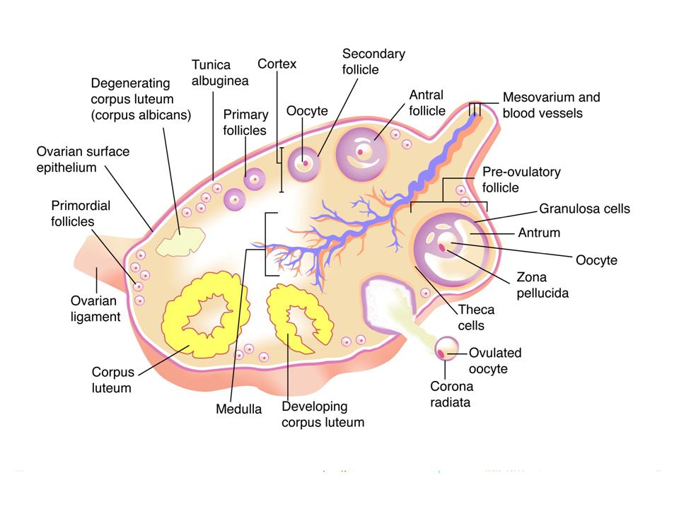
Source of Diagram 2 (above): Standard Class 12 Biology textbook illustration (CBSE/NCERT level – same as FSC syllabus).
Internal Structure of Ovary & Oogenesis
- Ovary shows different stages of follicular development.
- Oogenesis: Starts before birth.
- Oogonia (2n) → Primary oocyte (2n) → Secondary oocyte (n) + first polar body
- Secondary oocyte is released at ovulation.
- Only one ovum is produced from one primary oocyte (unlike 4 sperms in males).
- Follicles: Primordial → Primary → Secondary → Graafian (mature) follicle ruptures at ovulation → Corpus luteum (secretes progesterone).
Write a Note On Menstrual Cycle in Human Female
Definition The menstrual cycle is a series of cyclic changes in the ovary and uterus of a sexually mature female that occurs every 28 days (approximately). It prepares the female body for pregnancy. If fertilization does not occur, the cycle ends with shedding of the uterine lining (menstruation).
DurationAverage 28 days (can vary from 21–35 days).
Starts at puberty (menarche, 11–15 years) and continues till menopause (45–55 years).
Four Phases of Menstrual Cycle (Teach with Diagram 1)
- Menstrual Phase (Days 1–5)
- Shedding of endometrium (uterine lining) along with blood (menstruation).
- Ovary: No new follicle development yet.
- Hormone levels: Low estrogen and progesterone.
- Follicular Phase (Days 6–13)
- Primary follicle matures into Graafian follicle under FSH.
- Uterus: Endometrium starts regenerating and thickens.
- Estrogen level rises → stimulates endometrial growth.
- Ovulatory Phase (Day 14)
- Sudden surge of LH causes rupture of Graafian follicle.
- Secondary oocyte is released into fallopian tube (ovulation).
- This is the only day when fertilization is possible.
- Luteal Phase (Days 15–28)
- Ruptured follicle changes into corpus luteum.
- Corpus luteum secretes large amount of progesterone.
- Uterus: Endometrium becomes thick, vascular and secretory (ready for implantation).
- If no fertilization → corpus luteum degenerates → progesterone falls → new cycle starts.
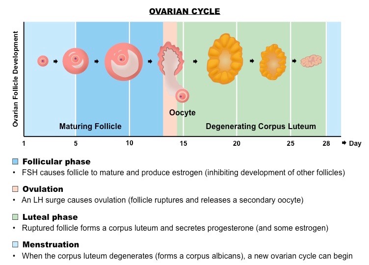
Hormonal Regulation (Very important for long questions – see Diagram 1)
- Hypothalamus releases GnRH.
- Pituitary releases FSH and LH.
- FSH → follicle development & estrogen secretion.
- LH surge → ovulation & corpus luteum formation.
- Estrogen → endometrial repair and growth.
- Progesterone → maintains thick endometrium.
If pregnancy occurs: hCG (from embryo) maintains corpus luteum.
If no pregnancy: Corpus luteum degenerates → menstruation.
Summary Table for Quick Revision
| Phase | Days | Ovarian Event | Uterine Event | Main Hormone |
|---|---|---|---|---|
| Menstrual | 1–5 | No activity | Endometrium sheds | Low all hormones |
| Follicular | 6–13 | Follicle matures | Endometrium regenerates | Estrogen ↑ |
| Ovulatory | 14 | Ovulation (oocyte released) | — | LH surge |
| Luteal | 15–28 | Corpus luteum forms | Endometrium thick & secretory | Progesterone ↑ |
Write a Detailed note on Birth
PARTURITION: THE PROCESS OF BIRTH
In humans, the process of giving birth is known as Parturition. According to the FSc Biology (Class 12) PTB curriculum, this process is controlled by a neuroendocrine mechanism involving both the mother and the fetus.
1. THE ONSET OF LABOR SIGNALS
The process begins when the fetus is fully developed. The following changes occur:
- Decrease in Progesterone: The level of progesterone, which maintains pregnancy by relaxing the uterus, drops significantly.
- Fetal ACTH & Corticosteroids: The fetal pituitary gland secretes ACTH, stimulating its adrenal glands to release corticosteroids. These cross the placenta and trigger the mother's body to start labor.
2. HORMONAL REGULATION (OXYTOCIN)
The most critical hormone for birth is Oxytocin, released by the mother’s Posterior Pituitary Gland.
- Contractions: Oxytocin stimulates the smooth muscles of the uterus (myometrium) to contract.
- Positive Feedback: As the baby's head pushes against the cervix, more oxytocin is released, leading to stronger and more frequent contractions known as Labor Pains.
3. STAGE I: CERVICAL DILATION
- The cervix (the opening of the uterus) softens and dilates to about 10 cm.
- The Amniotic Sac ruptures, and the protective amniotic fluid is discharged (often called "water breaking").
4. STAGE II: EXPULSION OF THE FETUS
- Powerful uterine contractions and abdominal pressure push the baby through the birth canal (vagina).
- The baby is delivered, and the Umbilical Cord is clamped and cut, marking the beginning of the infant's independent existence.
5. STAGE III: THE AFTERBIRTH (PLACENTAL STAGE)
- Within 10–45 minutes after the baby is born, the uterus contracts again.
- The Placenta and remaining fetal membranes detach from the uterine wall and are expelled from the mother’s body. This is known as the Afterbirth.
6. POST-NATAL ADAPTATIONS
- Lactation: The hormone Prolactin stimulates milk production in the mammary glands.
- Immunity: The first milk, called Colostrum, is rich in antibodies to protect the newborn from infections.
Write a Note On STD
Gonorrhea:
Gonorrhea is a common sexually transmitted infection caused by the bacterium Neisseria gonorrhoeae. It can affect both men and women and primarily targets the genital tract but can also infect the rectum, throat, and eyes.
Symptoms:
- Painful urination
- Abnormal discharge from the genital tract (often yellow or green in color)
- Pain during intercourse
- Swollen or painful testicles in men
- Sore throat (if transmitted orally)
In women, gonorrhea can lead to pelvic inflammatory disease (PID), which may cause infertility if left untreated. In men, it can lead to epididymitis, a painful condition of the reproductive system.
Treatment:
Gonorrhea is treatable with antibiotics. However, some strains have developed resistance to certain drugs, so a combination of antibiotics may be required.
Syphilis:
Syphilis is caused by the bacterium Treponema pallidum. It progresses in four stages: primary, secondary, latent, and tertiary. Early detection and treatment are essential to avoid severe health complications.
Stages of Syphilis:
-
Primary Stage: A painless sore (chancre) appears at the site of infection, usually around the genital, anal, or mouth area.
-
Secondary Stage: Skin rashes, mucous membrane lesions, and flu-like symptoms develop.
-
Latent Stage: The disease becomes dormant without symptoms but remains in the body. It can still be transmitted to others.
-
Tertiary Stage: If untreated, syphilis can lead to serious health problems like heart damage, brain damage, and other organ complications.
Symptoms:
- Painless sores in the genital area (primary stage)
- Skin rashes, mucous lesions (secondary stage)
- Swollen lymph nodes, fever
- No symptoms in the latent stage
Treatment:
Syphilis can be treated with antibiotics, especially penicillin, which is highly effective if administered during the early stages.
Herpes Simplex Virus (HSV):
Herpes Simplex Virus (HSV) causes both oral and genital herpes. There are two types of the virus:
- HSV-1: Primarily causes oral herpes, leading to cold sores around the mouth.
- HSV-2: Mostly causes genital herpes, resulting in painful sores in the genital area.
HSV is highly contagious and can be spread through direct contact with sores or even skin-to-skin contact when no visible sores are present.
Symptoms:
- Painful blisters or sores around the genital area, mouth, or anus
- Itching or discomfort in the affected area
- Flu-like symptoms, including fever and swollen lymph nodes (during the first outbreak)
- Recurring outbreaks, which may be less severe
Treatment:
There is no cure for herpes, but antiviral medications (e.g., acyclovir) can help manage symptoms, reduce the frequency of outbreaks, and lower the risk of transmission.
AIDS (Acquired Immunodeficiency Syndrome):
AIDS is the final stage of HIV (Human Immunodeficiency Virus) infection. HIV attacks the body's immune system, specifically the CD4 cells (T cells), weakening its ability to fight infections. If left untreated, HIV can progress to AIDS, making the person highly vulnerable to opportunistic infections and cancers.
Symptoms:
- Rapid weight loss
- Recurring fever or night sweats
- Extreme tiredness and fatigue
- Unexplained weight loss
- Swollen lymph nodes
- Frequent infections
Treatment:
Although there is no cure for HIV/AIDS, antiretroviral therapy (ART) is used to control the virus and prevent it from progressing to AIDS. ART helps lower the viral load to undetectable levels, enabling people with HIV to live long and healthy lives while reducing the risk of transmission to others.
Prevention:
- Safe sexual practices, such as using condoms, are crucial in preventing the spread of HIV.
- Regular HIV testing and early treatment can significantly improve outcomes.
Write a Note On Meristem
Types of Meristems
Meristems are regions of undifferentiated cells in plants that are capable of continuous division and growth. These tissues are responsible for the plant's growth in length and girth. There are three main types of meristems based on their location and function:
1. Apical Meristems
Apical meristems are found at the tips of roots and shoots. These meristems are responsible for primary growth, which results in an increase in the length of the plant. They are located at the apex (or growing tip) of roots and shoots and contribute to the development of new leaves, stems, and flowers.
Location:
- At the tip of stems (shoot apex)
- At the tip of roots (root apex)
Function:
- Elongation of roots and stems
- Formation of primary tissues like epidermis, cortex, vascular tissue, and pith
Subtypes:
- Shoot Apical Meristem (SAM): Found at the tips of shoots and is responsible for the growth of the shoot and formation of leaves.
- Root Apical Meristem (RAM): Located at the tips of roots and is responsible for the growth of the root.
2. Lateral Meristems
Lateral meristems are responsible for secondary growth, which leads to an increase in the girth or thickness of the plant. These meristems are found along the sides of stems and roots and contribute to the formation of woody tissues in dicots and gymnosperms.
Location:
-
In the lateral regions of stems and roots
Function:
- Increase in the diameter or girth of stems and roots
- Formation of secondary tissues, such as xylem and phloem
Subtypes:
- Vascular Cambium: Located between the xylem and phloem, it is responsible for the production of secondary xylem (wood) and secondary phloem (part of the bark).
- Cork Cambium (Phellogen): Produces the outer protective layers like cork and is responsible for the formation of the periderm (the outer bark in woody plants).
3. Intercalary Meristems
Intercalary meristems are present in some plants, primarily monocots, such as grasses. These meristems are located between mature tissues, often at the base of internodes or leaf blades. They contribute to the regrowth of leaves and stems and help the plant recover from damage or pruning.
Location:
- Found between mature tissues, such as in the base of leaves or internodes
Function:
- Allow for the elongation of internodes
- Facilitate regrowth after damage or cutting, especially in grasses and other monocots
Explain Apical Dominance
Apical Dominance
Apical dominance is a phenomenon in plants where the main central stem (or shoot) grows more vigorously than the lateral branches or shoots. This is a form of growth regulation that ensures the plant's growth is directed upwards, optimizing light capture and resources for overall survival and reproduction.
Apical dominance is primarily controlled by the apical meristem at the tip of the plant’s shoot. The apical meristem is the region of active cell division that is responsible for the growth of the plant's main stem. The dominance of the apical meristem over lateral buds (the small buds found along the sides of the stem) ensures that the plant grows taller and more rapidly in order to compete for light and space.
Mechanism of Apical Dominance:
The primary mechanism behind apical dominance involves the production of hormones, particularly auxins:
- Auxin Production: The shoot tip (apical meristem) produces auxin, a plant hormone that inhibits the growth of lateral (side) buds. Auxins move downward from the shoot tip to the rest of the plant, suppressing the growth of lateral buds.
- Lateral Bud Inhibition: As auxin levels in the lateral buds are high due to the apical meristem’s proximity, the growth of these buds is inhibited. This results in the lateral branches remaining dormant while the main stem continues to grow.
- Auxin Gradient: The further the lateral buds are from the apical meristem, the less auxin they receive, allowing them to grow. This gradient helps in the formation of a plant with a strong, upright central stem.
Role of Cytokinins:
Cytokinins are another class of plant hormones that promote cell division and growth in lateral buds. The ratio of auxins to cytokinins is critical in controlling apical dominance. If auxin production from the apical meristem is reduced, the influence of cytokinins on lateral bud growth increases, leading to the growth of lateral branches.
Factors That Affect Apical Dominance:
- Removal of the Apical Meristem: If the apical meristem (top bud) is removed (e.g., through pruning or damage), the inhibition of lateral buds decreases, and these buds are allowed to grow. This is why pruning often results in the growth of side branches.
- Light: Light availability can also affect apical dominance. In low light conditions, plants may exhibit less apical dominance and allow for more lateral growth to maximize light capture.
- Environmental Stress: Factors such as nutrient availability, water stress, and competition can influence apical dominance by affecting hormone levels within the plant.
Describe Role Of Nucleus In Developement
Role of Nucleus in Development
The nucleus is the control center of the cell, as it contains the cell's genetic material (DNA). It is essential for regulating cellular activities, including gene expression, cell division, and differentiation. During development, the nucleus controls the processes that lead to the formation of specialized cells, tissues, and organs.
Key Functions of the Nucleus in Development:
-
Genetic Information Storage: The nucleus stores the DNA, which contains the instructions necessary for cellular activities and development.
-
Regulation of Gene Expression: The nucleus controls the expression of genes, which directs the production of proteins responsible for cellular functions.
-
Cell Division: The nucleus is involved in both mitosis and meiosis, ensuring the accurate division of genetic material to daughter cells during cell division.
-
Cell Differentiation: During development, the nucleus helps direct the differentiation of stem cells into specialized cell types, such as muscle cells or nerve cells.
Hammerling’s Experiment: Nucleus and Development
Carl Hammerling conducted experiments in the 1930s to investigate the role of the nucleus in development using a type of green algae called Acetabularia. His work was pivotal in demonstrating that the nucleus plays a central role in the development and morphogenesis of organisms.
Objective of the Experiment:
Hammerling wanted to determine whether the nucleus contained the complete genetic information required for the development of the organism, specifically for the formation of its body shape and structure.
Hammerling’s Experiment:
Materials:
- Acetabularia (a single-celled green algae with a stalk and a cap-like structure)
- Microscope
- Scalpel for dissection
Procedure:
-
Isolation of the Nucleus:
-
Hammerling cut off the cap (the top part of the algae) from the stalk.
-
He then transplanted the nucleus from one type of Acetabularia to another (i.e., from one algae strain to another).
-
-
Nuclear Transplantation:
-
After removing the nucleus from one individual (which caused the algae to die or remain undeveloped), he transplanted the nucleus into another algae's stalk.
-
In some cases, Hammerling also performed cytoplasmic grafting, where he transplanted a nucleus from a different algae strain into a host, while keeping the rest of the algae's cytoplasm intact.
-
-
Observing Development:
-
Hammerling observed how the algae developed and what structures formed based on the donor nucleus. He specifically looked for the formation of caps on the algae stalks and whether the cap matched the traits of the donor.
-
Observations:
-
Cap Formation:
-
When Hammerling transplanted the nucleus of one strain of Acetabularia into another strain, the cap formation was determined by the nucleus rather than the surrounding cytoplasm. The cap grew in the shape characteristic of the algae strain from which the nucleus was taken, even though the cytoplasm was from a different strain.
-
-
Nucleus-Dependent Development:
-
If the nucleus was removed or replaced, the algae failed to produce the appropriate cap. This showed that the nucleus contained the genetic instructions required for the development of specific structures (such as the cap).
-
-
Cytoplasm Alone Did Not Control Development:
-
Even though the cytoplasm from the host algae was used, it did not control the final development of the organism. This experiment confirmed that the nucleus was responsible for determining the organism’s structural development, rather than the cytoplasm alone.
write a Note On Aging?
Aging refers to the gradual process of physical and biological changes that occur in an organism over time, leading to a decline in its functionality and vitality. It is a complex phenomenon involving multiple genetic, environmental, and lifestyle factors. Aging is characterized by the deterioration of cellular structures and the loss of biological functions, ultimately leading to an increased vulnerability to diseases and death.
Key Concepts in Aging:
-
Cellular Aging:
-
As organisms age, their cells experience various forms of damage due to environmental factors (such as UV radiation and pollutants), metabolic processes (free radical damage), and errors during cell division.
-
Telomere Shortening: The telomeres, protective caps at the ends of chromosomes, shorten with each cell division. Eventually, the telomeres become too short for the cell to divide, leading to cell death or senescence (a state of permanent growth arrest).
-
Accumulation of Cellular Damage: Damage from free radicals, oxidative stress, and DNA mutations accumulates over time, impairing cellular function.
-
-
Genetic Theories of Aging:
-
Programmed Theories: These theories suggest that aging is genetically programmed and governed by the body’s biological clock. Certain genes are believed to regulate the lifespan of an organism, including genes that control cell division and repair mechanisms.
-
Damage or Error Theories: These theories propose that aging occurs due to the accumulation of random molecular damage over time, such as DNA mutations, protein damage, and oxidation of cellular components. This damage disrupts cellular processes and leads to aging.
-
-
Physiological Changes During Aging:
-
Skin: The skin loses elasticity and thickness, leading to wrinkles and sagging. The rate of skin cell turnover decreases, and collagen production slows down.
-
Muscle Mass and Strength: There is a decline in muscle mass and strength, a condition known as sarcopenia. This is partly due to the loss of muscle fibers and reduced protein synthesis.
-
Bone Density: Bone mass decreases with age, leading to conditions such as osteoporosis, making bones more fragile and susceptible to fractures.
-
Cardiovascular System: The heart becomes less efficient in pumping blood, and blood vessels lose elasticity, contributing to increased blood pressure and the risk of heart disease.
-
Immune System: The immune system weakens with age, leading to increased susceptibility to infections and diseases. This decline in immune function is referred to as immunosenescence.
-
Cognitive Function: Aging affects the brain, leading to slower cognitive processing, memory decline, and in some cases, neurodegenerative diseases like Alzheimer’s disease.
-
-
Hormonal Changes:
-
As individuals age, there is a decrease in the production of key hormones such as growth hormone, estrogen, and testosterone. These hormonal changes contribute to the physical and metabolic changes seen in aging individuals.
-
Factors Affecting Aging:
-
Genetics:
-
Genetic factors play a significant role in determining the rate and extent of aging. Some individuals inherit genes that promote longevity, while others may inherit genes that predispose them to age-related diseases.
-
-
Environmental Factors:
-
Exposure to Sunlight: Overexposure to ultraviolet (UV) radiation from the sun accelerates the aging process, particularly by damaging the skin and increasing the risk of skin cancer.
-
Pollution and Toxins: Environmental pollutants and toxins can contribute to the aging process by increasing oxidative stress and DNA damage.
-
Diet and Lifestyle: A balanced diet rich in antioxidants, regular physical activity, and avoiding smoking and excessive alcohol consumption can slow down the aging process and improve overall health.
-
-
Free Radical Theory of Aging:
-
According to this theory, free radicals—unstable molecules that are produced during normal metabolic processes—cause oxidative damage to cells. Over time, this damage accumulates and contributes to aging.
-
-
Telomere Theory:
-
This theory suggests that the shortening of telomeres during cell division is a key factor in aging. Once telomeres become too short, cells can no longer divide, leading to tissue degeneration and aging.
-
Aging and Disease:
-
Aging is closely linked to an increased risk of developing various diseases, including cardiovascular diseases, cancer, diabetes, and neurodegenerative diseases like Parkinson’s and Alzheimer’s disease.
-
Chronic Inflammation: Chronic low-level inflammation, often referred to as inflammaging, is a hallmark of aging and is associated with many age-related diseases.
Extending Lifespan:
While aging cannot be completely stopped, certain practices may help slow the process and improve the quality of life in older individuals:
-
Healthy Diet:
-
A diet rich in fruits, vegetables, whole grains, lean proteins, and healthy fats can help reduce oxidative stress, inflammation, and cellular damage.
-
Caloric Restriction: Studies in animals suggest that reducing calorie intake without malnutrition can extend lifespan and delay the onset of age-related diseases.
-
-
Physical Activity:
-
Regular exercise improves cardiovascular health, muscle strength, and bone density. It also helps maintain cognitive function and reduces the risk of chronic diseases.
-
-
Mental Health and Cognitive Stimulation:
-
Staying mentally active through reading, puzzles, learning new skills, and socializing can help preserve cognitive function and prevent age-related decline.
-
-
Anti-Aging Therapies:
-
Research in anti-aging therapies includes drugs, supplements, and interventions aimed at slowing the aging process. For example, metformin and rapamycin have been studied for their potential to extend lifespan in animal models.
what is regeneration explain with different examples
Regeneration
Regeneration is the process by which organisms can regrow lost or damaged body parts or tissues. This ability is common in certain organisms, and it varies across species. It involves the activation of undifferentiated cells (such as stem cells), which then proliferate and differentiate to form new, functional tissues or body parts.
Regeneration plays a vital role in the survival of many organisms, enabling them to recover from injuries and continue normal functions. The extent and efficiency of regeneration differ among species, with some organisms capable of regenerating entire organs or even complete bodies.
Types of Regeneration
-
Complete Regeneration:
-
This involves the regrowth of an entire body part or organ that was lost or damaged. The new tissue is fully functional and identical to the original tissue.
-
-
Partial Regeneration:
-
In this type, only part of the lost or damaged body part is regrown. The regenerated tissue may not function as effectively as the original, and in some cases, it may not regrow at all.
-
-
Morphallaxis and Epimorphosis:
-
Morphallaxis: In this form of regeneration, the remaining tissue reorganizes and reshapes to form the missing part.
-
Epimorphosis: The lost body part is regrown by the proliferation and differentiation of stem cells into the required structure.
-
Examples of Regeneration in Different Organisms
-
Regeneration in Starfish (Sea Stars):
-
Starfish are well-known for their ability to regenerate lost limbs. When a limb is lost (due to predation or injury), the starfish can regenerate the missing part, including the central disc. In some cases, a single arm can regenerate a whole new starfish if part of the central disc is present.
Mechanism:
The process involves the activation of stem cells, which divide and differentiate to form new tissues like the epidermis, muscle, and even the central disc. -
-
Regeneration in Planarians:
-
Planarians, a type of flatworm, are remarkable for their regenerative abilities. If a planarian is cut into pieces, each piece can regenerate into a fully functional individual.
Mechanism:
Planarians have a large number of undifferentiated stem cells called neoblasts that can differentiate into any cell type, allowing them to regenerate missing body parts, including the head and tail. -
-
Regeneration in Salamanders:
-
Salamanders, especially species like the axolotl, are famous for their ability to regenerate lost limbs, tails, heart tissue, and even parts of the spinal cord.
Mechanism:
After amputation, specialized cells at the wound site form a blastema, a mass of proliferating cells that will differentiate to form the lost tissues. The process is controlled by a complex network of molecular signals, including growth factors and proteins. -
-
Regeneration in Lizards:
-
Some lizards, such as the gecko, can regenerate their tails if they are lost due to predation or injury. The regenerated tail may not be identical to the original, but it serves a similar function in balance and movement.
Mechanism:
After tail loss, cells at the wound site undergo rapid proliferation, forming a new tail. The new tail is often made of cartilage rather than the bone found in the original tail. -
-
Regeneration in Hydra:
-
Hydra, a small freshwater organism, has an extraordinary regenerative capacity. Even if a small piece of a hydra is cut off, it can regenerate into a complete, functional organism.
Mechanism:
Hydra possesses abundant stem cells, which proliferate and differentiate into the various tissues necessary for regeneration, including the body column, tentacles, and mouth. -
-
Regeneration in Humans (Limited):
-
Humans have a limited ability to regenerate certain tissues. For example:
-
The liver has a remarkable regenerative capacity. If part of the liver is removed, the remaining part can grow back to nearly its original size.
-
Skin and bone cells also exhibit regenerative abilities. When injured, the skin regenerates through cell division, and bone cells repair fractures by regenerating bone tissue.
-
However, humans are not capable of regenerating complex structures like limbs or organs such as the heart.
-
Mechanisms of Regeneration
-
Stem Cells:
-
Regeneration often involves the activation of stem cells—undifferentiated cells that can divide and develop into specialized cell types. These cells proliferate to replace lost or damaged tissues.
-
-
Blastema Formation:
-
In animals like salamanders, a group of undifferentiated cells forms a blastema at the site of injury. The blastema then differentiates into the necessary cell types (e.g., muscle, nerve, bone) to restore the lost part.
-
-
Gene Expression and Growth Factors:
-
During regeneration, specific genes are activated that are usually silent in the adult organism. Growth factors such as FGF (Fibroblast Growth Factor) and BMPs (Bone Morphogenetic Proteins) play a significant role in signaling cells to divide and differentiate.
-
Write a Note On Abnormal Develoment
Abnormal Development
Abnormal development occurs when the normal process of development is disturbed, leading to defects or malformations in the organism. These abnormalities can arise due to various factors such as genetic mutations, environmental influences, and metabolic defects.
Causes of Abnormal Development:
-
Genetic Abnormalities:
-
Inheritance of Defective Genes: Abnormalities may be inherited from parents due to defective or abnormal genes. The inheritance may depend on whether the gene is dominant or recessive, homozygous or heterozygous.
-
Sex Chromosome Abnormalities: Abnormalities can also occur due to the presence of an extra or missing sex chromosome, leading to syndromes:
-
Klinefelter's Syndrome (XXY): A trisomy of the sex chromosomes, leading to tallness, aggressiveness, mental defects, and antisocial behavior.
-
Turner's Syndrome (XO): A condition where one sex chromosome is missing, leading to various developmental issues, including short stature and infertility.
-
-
-
Environmental Factors (Teratogens):
-
Teratogens are environmental agents that cause developmental abnormalities. These include:
-
- Ionizing Radiation (e.g., X-rays): These can affect the developing egg or sperm and cause mutations in the genes.
- Nutritional Deficiencies: Lack of essential substances like vitamins and trace elements can hinder the differentiation of cells in the fetus.
- Toxins and Drugs: Substances ingested by the mother, such as drugs or toxins, can disrupt fetal development, potentially leading to malformations.
-
Metabolic Defects:
-
Metabolic defects can cause structural deviations during organogenesis (the formation of organs). These defects may result in the absence or repetition of body parts.
- Microcephaly: A condition where individuals are born with a small skull.
- Cleft Palate and Harelip: Abnormal development of facial features.
- Polydactyly or Syndactyly: Conditions where the number of fingers or toes is more or less than normal.
- Density-Dependent and Density-Independent Factors: Mechanisms Influencing Population Dynamics
- MDCAT Past Papers with Most Repeated MCQs (2025 Preparation)
- Exploring the Types and Physiological Mechanisms of Seed Germination in Plants


.jpg)


4 Comments
Definitely and totally cured of herpes virus by Doctor Razor's Quick cure for Herpes. My Sincere Gratitude to Dr Razor, I was infected with HERPES SIMPLEX VIRUS 2 November 13 2018, I went to many hospitals for cure but there was no solution, I was confused because the medical drugs has become my daily food, but i decided to sort after Natural Herbs. One day I sat beside the pool, Browsing and thinking where I could get a solution to end this predicament. I saw a blog on how Herbalist Razor cured people with his herbal medicine, I decided to Visit this Doctor's Razor Website https://herbalistrazorherb.wixsite.com/drrazorherbalhome and being satisfied, I contacted and Explained my Problems to him and he prepared the herbs for me which I took for a period of time and he instructed me to go for a check up, after the test I was confirmed herpes negative, MY JOY KNOWS NO BOUND. This testimony serves as a token of my gratitude.He renders Treatment for :
ReplyDeleteHIV/AIDS,
SHINGLES,
CANCER,
HUMAN PAPILLOMA VIRUS,
ASTHMA,
DIABETES
BARRENNESS/INFERTILITY P*NIS ENLARGEMENTSPELL COUNSELING.
You Can decide to reach Herbalist Razor on his Email address : drrazorherbalhome@gmail.com . Cell Phone/Whatsapp Number +2349065420442
thank you so much mam for nice informations
Deletethanks for email this will really help many peoples .God bless you.
Deletethanks to you too if you think so...
Delete