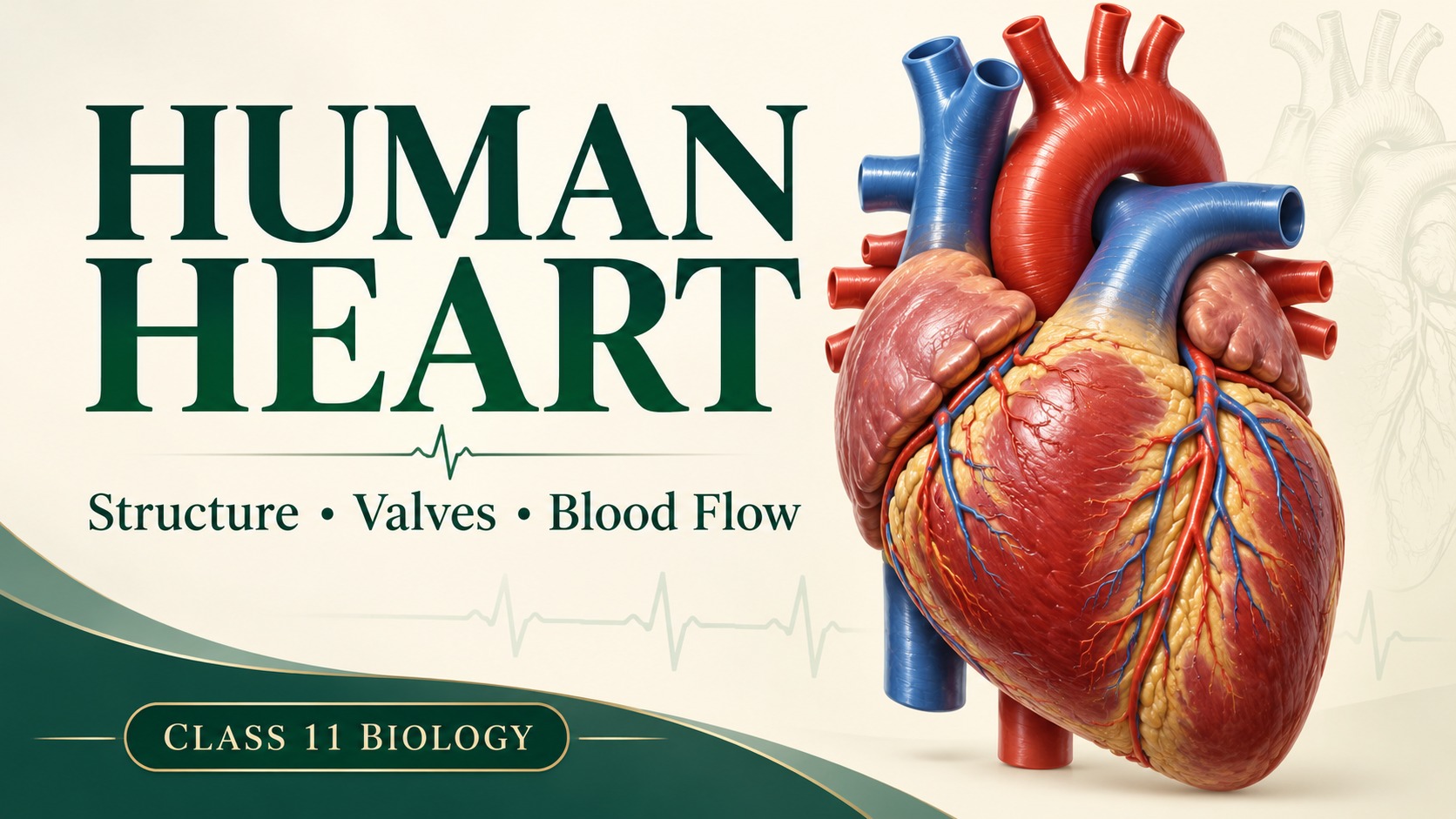
Understand each heart structure by the job it performs, then trace a complete blood route without guessing.
1. Start with the job: two pumps working together
A useful way to understand the human heart is to follow one journey of blood. The right side sends blood to the lungs, where gas exchange takes place. The left side sends blood into systemic circulation to supply the body's tissues. The two sides work together, but they maintain separate routes in the normal adult heart. (Think of two coordinated pumps in one organ, not one chamber with four exits.)
The heart creates pressure differences that move blood. Its muscular walls provide the force, while its valves maintain the direction. These roles are different: a valve does not squeeze blood forward, and a strong muscular wall alone cannot prevent backflow. This structure–function relationship will explain nearly every feature below.
2. Where is the heart? Use anatomical right and left
The heart lies in the thoracic cavity between the lungs, behind the sternum. Much of it lies slightly to the left of the midline, and its apex points downwards and towards the left. It is therefore misleading to imagine the entire heart sitting inside the left side of the chest.
Right and left always refer to the person's own sides. In a diagram viewed from the front, the person's right side appears on the viewer's left. (Before labelling a heart, find the right atrium and the thick-walled left ventricle; do not assign sides using your own hands.)
3. Pericardium: a protective sac with a lubricated space
The pericardium encloses the heart. Its outer fibrous component is tough connective tissue that helps hold the heart in place and limits excessive expansion. The serous pericardium has a parietal layer lining the inside of the fibrous sac and a visceral layer closely covering the heart. The visceral serous layer is also called the epicardium.
The narrow pericardial cavity lies between the parietal and visceral serous layers. Its small amount of fluid reduces friction as the heart repeatedly moves. (Two surfaces can slide more smoothly when a thin lubricating film separates them.) Keep the cavity outside the heart wall in your mental picture; it is not an extra blood-filled chamber.
4. The heart wall: outside, muscle, inside
The wall has three main layers. Epicardium is the outer covering. Myocardium is the muscular middle layer and produces the force of contraction. Endocardium forms the smooth inner lining of the chambers and covers the valve surfaces. A useful order is E–M–E: epicardium, myocardium, endocardium, from outside to inside.
Do not equate the entire pericardium with one layer of the heart wall. The visceral serous pericardium is the epicardium, but the surrounding fibrous sac and parietal layer are not myocardium. Similarly, myocardium is cardiac muscle; it is not the same as endocardium simply because both names contain 'cardium'.
5. Four chambers: receiving above, pumping below
The right and left atria are the upper receiving chambers. The right atrium receives blood returning from the systemic circuit, chiefly through the superior and inferior venae cavae. The left atrium receives blood returning from the lungs through the pulmonary veins. Atria pass blood into their corresponding ventricles.
The right and left ventricles are the lower pumping chambers. The right ventricle ejects blood into the pulmonary trunk, which divides into pulmonary arteries. The left ventricle ejects blood into the aorta. Atria have relatively thin muscular walls because they move blood over the short distance into the ventricles. Ventricles must generate the pressure needed to move blood out into their circuits.
6. Why is the left ventricular wall thicker?
The left ventricle has a much thicker myocardium than the right ventricle because the systemic circuit requires a higher pressure. The right ventricle supplies the lower-pressure pulmonary circuit. (The key link is workload and resistance, not that oxygenated blood is heavier or that the left ventricle stores a larger batch of blood.)
In steady circulation, both ventricles pump approximately the same volume over time. A thicker left ventricular wall therefore means greater pressure-generating capacity, not permission to pump several times as much blood as the right ventricle. This distinction prevents a common error in comparison answers.
7. Septa: keeping the routes separate
The interatrial septum separates the two atria; the interventricular septum separates the two ventricles. These partitions help prevent mixing between the right and left sides in the normal adult heart. The interventricular septum is muscular and thicker than the interatrial septum.
A septum is a separating wall, whereas a valve is a movable structure guarding an opening. Do not draw a valve between the right and left ventricles. Blood normally travels from a right-sided chamber to a left-sided chamber by passing through the pulmonary circuit, not through an opening in the septum.
8. Four valves: learn the doorway and the direction
The tricuspid valve lies between the right atrium and right ventricle. The bicuspid or mitral valve lies between the left atrium and left ventricle. Together they are atrioventricular (AV) valves. During ventricular contraction, they prevent blood returning into the atria. Their names refer to three and two principal cusps respectively.
The pulmonary semilunar valve guards the exit from the right ventricle into the pulmonary trunk. The aortic semilunar valve guards the exit from the left ventricle into the aorta. They prevent blood returning from these arteries into the relaxing ventricles. (AV valves protect the inlet; semilunar valves protect the outlet.)
Valves open and close in response to pressure differences across them. They do not actively decide when to open. Follow the pressure on each side, then predict the permitted direction. The timing of these changes is explained in the linked cardiac-cycle lesson.
Heart valves at a glance
| Valve | Location | Prevents backflow into |
|---|---|---|
| Tricuspid | Right atrium → right ventricle | Right atrium |
| Mitral / bicuspid | Left atrium → left ventricle | Left atrium |
| Pulmonary semilunar | Right ventricle → pulmonary trunk | Right ventricle |
| Aortic semilunar | Left ventricle → aorta | Left ventricle |
9. Trace one complete blood pathway
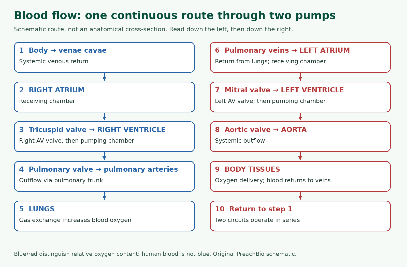
Use this ordered route: body → venae cavae → right atrium → tricuspid valve → right ventricle → pulmonary valve → pulmonary trunk and arteries → lungs → pulmonary veins → left atrium → mitral valve → left ventricle → aortic valve → aorta → body. Say the route aloud once, then redraw it without looking.
Pulmonary circulation connects the right ventricle to the lungs and back to the left atrium. Systemic circulation connects the left ventricle to the body's tissues and back to the right atrium. Together these make double circulation: blood passes through the heart twice during a complete journey through both circuits. (Do not interpret 'double' as two passes through the same ventricle.)
10. Artery and vein: direction is the rule
An artery carries blood away from the heart; a vein carries blood towards it. In the adult pulmonary circuit, the pulmonary arteries carry relatively deoxygenated blood, while the pulmonary veins carry oxygenated blood. These are not exceptions to the direction rule. They are exceptions only to the mistaken claim that all arteries carry oxygenated blood.
Deoxygenated does not mean absolutely oxygen-free. It means the blood has a lower oxygen content than oxygenated blood in this comparison. Blue in teaching diagrams is a convention for distinguishing routes; human blood is not blue. A good answer separates the colour key from the biological meaning.
11. Worked reasoning: identify a chamber without memorising its position
Question: a chamber receives blood through the mitral valve and ejects it through an aortic valve. Which chamber is it? First identify the inlet: the mitral valve leads from the left atrium to the left ventricle. Then check the outlet: the aorta leaves the left ventricle. Both clues agree, so the chamber is the left ventricle.
Question: why would closing the tricuspid valve be useful when the right ventricle contracts? Ventricular pressure rises above atrial pressure. Without a competent valve, some blood could move backwards into the right atrium instead of continuing towards the pulmonary trunk. This answer links location, pressure and function rather than repeating 'prevents backflow' without naming its direction.
12. Build an exam answer that explains, not just lists
For a structure-and-function answer, organise your response under position and coverings, wall layers, chambers and septa, valves, then the blood pathway. Pair each structure with its purpose. For example, 'The left ventricle has a thick myocardium that generates the pressure required for systemic circulation' is stronger than 'The left ventricle is thick.'
For a diagram answer, use clear leader lines, label anatomical sides correctly, show the septum, and name the four valves at their proper openings. Finish by checking that pulmonary veins enter the left atrium and the aorta leaves the left ventricle. Those two checkpoints catch many otherwise attractive but incorrect drawings.
20 MCQs with explained answers
Attempt each question before opening its explanation. All questions are original practice for this lesson.
1. Which chamber normally receives the venae cavae?
- Left ventricle
- Left atrium
- Right ventricle
- Right atrium
Show answer and explanation
D. Right atrium — Systemic venous return enters the right atrium before passing into the right ventricle.
2. Which valve connects the left atrium to the left ventricle?
- Mitral valve
- Aortic valve
- Pulmonary valve
- Tricuspid valve
Show answer and explanation
A. Mitral valve — Mitral and bicuspid name the left AV valve.
3. Which heart-wall layer produces most of the contraction force?
- Pericardial fluid
- Parietal pericardium
- Myocardium
- Endocardium
Show answer and explanation
C. Myocardium — Myocardium consists of cardiac muscle.
4. Where is the pericardial cavity?
- Between the atria
- Between parietal and visceral serous layers
- Inside the aorta
- Between the ventricles
Show answer and explanation
B. Between parietal and visceral serous layers — It is a lubricated space outside the heart wall, not a blood chamber.
5. What is the main function of pericardial fluid?
- Reduce friction during heart movement
- Carry oxygen to the left atrium
- Close the aortic valve
- Trigger the SA node
Show answer and explanation
A. Reduce friction during heart movement — Lubrication reduces friction between moving serous surfaces.
6. Which structure is also called epicardium?
- Interatrial septum
- Visceral serous pericardium
- Endocardium
- Fibrous pericardium
Show answer and explanation
B. Visceral serous pericardium — The visceral serous layer lies directly on the heart.
7. Why is the left ventricular myocardium thicker?
- It receives the venae cavae
- It contains heavier blood
- It generates the pressure required for systemic flow
- It pumps several times the right ventricular volume
Show answer and explanation
C. It generates the pressure required for systemic flow — Greater systemic pressure workload requires a thicker muscular wall.
8. Which vessel directly receives blood from the right ventricle?
- Superior vena cava
- Pulmonary vein
- Pulmonary trunk
- Aorta
Show answer and explanation
C. Pulmonary trunk — The right ventricle ejects through the pulmonary valve into the pulmonary trunk.
9. Which vessel returns oxygenated blood from the lungs?
- Pulmonary trunk
- Inferior vena cava
- Pulmonary vein
- Pulmonary artery
Show answer and explanation
C. Pulmonary vein — Pulmonary veins enter the left atrium after pulmonary gas exchange.
10. What separates the two ventricles?
- Pericardial cavity
- Mitral valve
- Interventricular septum
- Interatrial septum
Show answer and explanation
C. Interventricular septum — The interventricular septum is the partition between ventricles.
11. Which valve prevents aortic blood returning to the left ventricle?
- Mitral valve
- Tricuspid valve
- Aortic semilunar valve
- Pulmonary semilunar valve
Show answer and explanation
C. Aortic semilunar valve — It guards the left ventricular outlet to the aorta.
12. An artery is best defined as a vessel that:
- Always contains deoxygenated blood
- Enters an atrium
- Always contains oxygenated blood
- Carries blood away from the heart
Show answer and explanation
D. Carries blood away from the heart — Direction relative to the heart defines an artery.
13. Which sequence is correct?
- Right ventricle → aortic valve → aorta
- Right atrium → mitral valve → left ventricle
- Left atrium → tricuspid valve → right ventricle
- Right atrium → tricuspid valve → right ventricle
Show answer and explanation
D. Right atrium → tricuspid valve → right ventricle — The tricuspid valve is the right AV valve.
14. Which two valves are atrioventricular valves?
- Mitral and aortic
- Tricuspid and mitral
- Tricuspid and pulmonary
- Aortic and pulmonary
Show answer and explanation
B. Tricuspid and mitral — AV valves lie between atria and their corresponding ventricles.
15. What directly makes a normal valve open or close?
- Blood oxygen content alone
- Pressure difference across it
- A separate skeletal muscle in each cusp
- The colour used in a diagram
Show answer and explanation
B. Pressure difference across it — Valves respond passively to changing pressure differences.
16. Why are atrial walls thinner than ventricular walls?
- They never contract
- They move blood into adjacent ventricles at lower pressure
- They contain no muscle
- They pump directly through the entire systemic circuit
Show answer and explanation
B. They move blood into adjacent ventricles at lower pressure — Atria have a less demanding pressure workload.
17. Which statement describes deoxygenated blood accurately?
- It is naturally blue
- It never contains carbon dioxide
- It can occur only in veins
- It has relatively less oxygen, not necessarily none
Show answer and explanation
D. It has relatively less oxygen, not necessarily none — Deoxygenated is a relative oxygen-content description.
18. Which normal adult route takes blood from the right to the left side of the heart?
- Through the pericardial cavity
- Through the lungs
- Across the interventricular septum
- Directly through the mitral valve from the right ventricle
Show answer and explanation
B. Through the lungs — The pulmonary circuit connects the right ventricular outlet to the left atrial inlet.
19. Which is the correct outside-to-inside order of heart-wall layers?
- Endocardium → epicardium → myocardium
- Myocardium → endocardium → epicardium
- Epicardium → myocardium → endocardium
- Epicardium → endocardium → myocardium
Show answer and explanation
C. Epicardium → myocardium → endocardium — Muscular myocardium lies between the outer covering and inner lining.
20. What does double circulation mean?
- The heart has two chambers
- Blood always crosses the same valve twice per heartbeat
- The left ventricle pumps twice the right ventricular volume
- Blood passes through the heart twice per complete pulmonary-and-systemic circuit
Show answer and explanation
D. Blood passes through the heart twice per complete pulmonary-and-systemic circuit — The pulmonary and systemic circuits are linked through the two sides of the heart.
Interactive quiz: 20 different questions
This is a separate question set. Choose one answer for each question, then check your score. No login, timer or personal information is needed; answers are processed in this page.
Quiz answer key and explanations — open after attempting
1: A. Left atrium — Pulmonary veins return blood from the lungs into the left atrium.
2: D. Left ventricle — These two valves border the left ventricle.
3: A. Fibrous pericardium — The tough fibrous component supports and restrains the heart.
4: C. Endocardium — Endocardium forms the internal chamber lining.
5: D. Interatrial septum — The atria are the receiving chambers.
6: C. Backflow into the right atrium — The right AV valve prevents ventricular-to-atrial backflow.
7: D. Pulmonary trunk — The pulmonary valve guards the right ventricular outlet.
8: A. Aorta — The aortic valve leads from the left ventricle into the aorta.
9: C. It carries blood away from the heart — The direction of flow makes it an artery despite its lower oxygen content.
10: C. Viewer's left — Anatomical sides belong to the subject being viewed.
11: B. Right ventricle—pulmonary circuit — The right ventricle pumps towards the lungs.
12: D. Pulmonary vein—right atrium — Pulmonary veins enter the left atrium, not the right atrium.
13: A. The two circuits operate in series — A sustained mismatch would accumulate blood in one part of the linked circulation.
14: C. The pericardial cavity receives systemic venous blood — Systemic venous blood enters the right atrium, not the surrounding serous cavity.
15: A. Two principal cusps — Bicuspid describes the valve's two principal leaflets.
16: C. Aorta — The aorta distributes left ventricular output to the systemic circulation.
17: C. Systemic veins → venae cavae → right atrium — The venae cavae provide the principal systemic venous entry to the right atrium.
18: A. A colour convention for relatively deoxygenated blood — The convention distinguishes routes; real blood is not blue.
19: B. Separation of right and left ventricular blood — The septum prevents direct passage between the ventricular cavities.
20: D. Thick left myocardium produces high systemic pressure — A strong answer identifies the mechanical requirement satisfied by the structure.
Student FAQs
Why is the left ventricle thicker than the right?
It generates the higher pressure required for systemic circulation. Both ventricles normally move approximately equal volumes over time; the difference is mainly pressure workload.
Is the heart entirely on the left side of the chest?
No. It lies between the lungs near the midline, with much of its mass and its apex directed towards the left.
What is the difference between pericardium and epicardium?
Pericardium is the enclosing sac with fibrous and serous components. Epicardium is its visceral serous layer covering the heart and is also the outer layer of the heart wall.
Are bicuspid and mitral valves the same?
Yes. Both names identify the left atrioventricular valve, between the left atrium and left ventricle.
Why does the pulmonary artery carry deoxygenated blood?
It carries blood from the right ventricle towards the lungs before that blood is oxygenated. Arteries are named by flow away from the heart.
Which valves stop blood entering the atria backwards?
The tricuspid and mitral AV valves prevent ventricular-to-atrial backflow when the ventricles contract.
Does deoxygenated blood contain no oxygen?
No. It contains less oxygen than oxygenated blood in this comparison, rather than necessarily containing none.
How can I memorise blood flow through the heart?
Learn right side → lungs → left side → body, then insert the receiving atrium, pumping ventricle and correctly positioned valves in each half.
Continue studying
- Cardiac cycle, heart sounds and pacemaker — apply the structures to one heartbeat.
- Homeostasis and feedback — related physiology revision at Class 12 level.
- Punjab Biology course resources and official textbook links
Sources, scope and preparation
Prepared for PreachBio on 20 October 2026. Original explanations, invented worked examples, questions and diagrams were developed for this lesson. Punjab textbook headings and section boundaries were checked; supplementary sources were used to clarify scientific wording. This is an independent learning resource, not an official PECTAA publication or medical advice.
- PECTAA official curriculum and textbook directory — follow the edition prescribed by your college.
- Punjab Biology 11, Chapter 11, section 11.1; printed pages 232–234. Public textbook copy consulted for curriculum scope.
- OpenStax: cardiac cycle — pressure changes and phase transitions.


0 Comments