Follow pressure, valves and electrical coordination to explain every stage of a heartbeat.
1. One heartbeat is a sequence, not one squeeze
A cardiac cycle is one complete sequence of contraction and relaxation of the heart chambers. Systole means contraction; diastole means relaxation. Atria and ventricles each have their own systole and diastole, so always name the chamber group you mean. (During ventricular systole, the atria are relaxing; 'the whole heart contracts' loses the essential coordination.)
The cycle is easiest to understand using three linked questions: which chambers are contracting, which side of each valve has greater pressure, and where can blood flow? Learn the sequence through those questions rather than memorising a list of open and closed valves without a reason. Read the linked heart-structure lesson first if you cannot yet trace the blood route.
2. The rule behind every valve movement
Blood tends to move from higher pressure to lower pressure. When atrial pressure exceeds ventricular pressure, an AV valve can open and permit filling. When ventricular pressure rises above atrial pressure, the AV valve closes. Semilunar valves open only when ventricular pressure exceeds the pressure in the corresponding outlet artery.
The heart muscle generates changing pressure; the valves respond passively. (A valve is more like a one-way flap than a motor.) This also explains why ventricular contraction does not immediately mean arterial ejection: pressure must rise enough to open the outlet valve first.
3. Stage 1: joint diastole allows filling
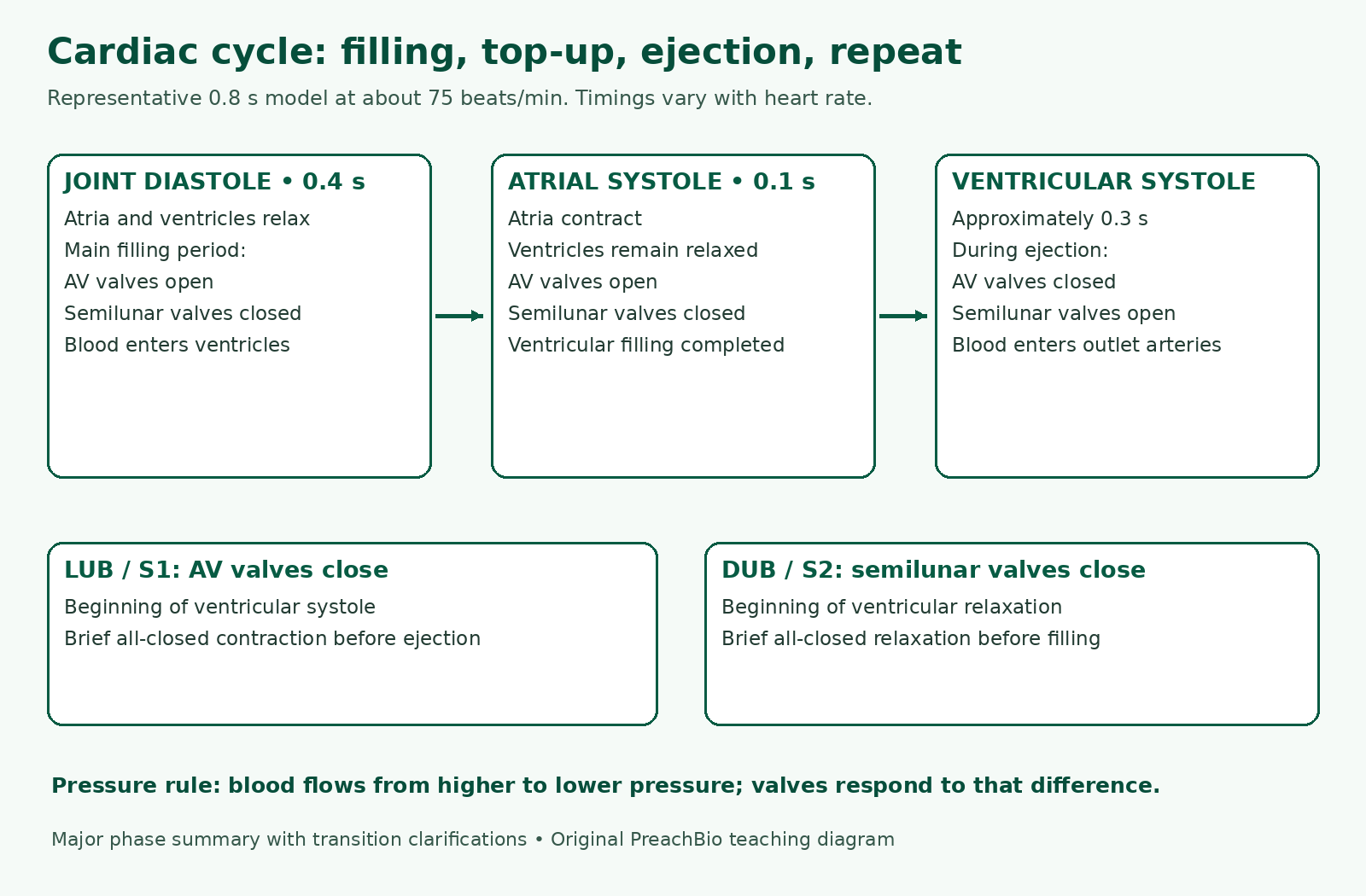
During the main shared relaxation period, both atria and ventricles are in diastole. Blood returns to the right atrium from systemic veins and to the left atrium from pulmonary veins. As the ventricles relax sufficiently, atrial pressure exceeds ventricular pressure, so the AV valves open and blood flows into the ventricles.
The semilunar valves remain closed because arterial pressure is greater than the pressure in the relaxing ventricles. Much ventricular filling occurs passively during relaxation. (Atria are not sealed storage tanks that wait to become completely full before releasing any blood.) There is a short transition immediately after ventricular contraction when all valves are closed; the filling description applies after AV valves open.
4. Stage 2: atrial systole tops up the ventricles
An electrical impulse initiated by the SA node spreads through the atria, causing atrial contraction. Atrial pressure rises and additional blood passes through the open AV valves into the relaxed ventricles. The semilunar valves remain closed. This stage completes ventricular filling before the ventricles contract.
It is incorrect to say rising atrial blood pressure itself is the normal pacemaker that triggers atrial systole. The normal rhythm begins with specialised cardiac muscle cells producing an electrical impulse. Pressure explains movement of blood; electrical excitation explains the coordinated initiation of contraction. Keep those two causes separate.
5. Stage 3: ventricular systole builds pressure, then ejects blood
When the ventricles contract, their pressure rises. The AV valves close as ventricular pressure exceeds atrial pressure. For a brief initial period, the semilunar valves have not yet opened either, so all four valves are closed. (This is a clarification of the transition: pressure can rise before blood leaves.)
Once ventricular pressure exceeds pulmonary-trunk or aortic pressure, the corresponding semilunar valve opens and ejection occurs. The right ventricle sends blood to the pulmonary trunk; the left sends blood to the aorta. During this ejection phase, AV valves are closed and semilunar valves are open. Both ventricles contract in a coordinated manner, but the left develops the higher pressure.
6. Return to diastole: outlet valves close first
As the ventricles relax, their pressure falls below the pressure in the outlet arteries. A brief tendency for blood to return closes the semilunar valves, preventing arterial backflow into the ventricles. Initially the AV valves are still closed. When ventricular pressure falls below atrial pressure, they open and filling resumes.
This brief all-closed relaxation interval is why a three-stage school diagram should not be interpreted as a frame-by-frame account of every instant. For the prescribed sequence, remember filling → atrial top-up → ventricular ejection → relaxation. Use the pressure rule whenever a question asks about a boundary between stages.
7. Timing: why 0.8 seconds means 75 beats per minute
The textbook's representative resting cycle uses approximately 0.1 s for atrial systole, 0.3 s for ventricular systole and 0.4 s for the shared relaxation period. These three successive teaching phases total 0.8 s. Heart rate = 60 ÷ cycle duration in seconds, so 60 ÷ 0.8 = 75 beats per minute.
These values are approximate, not fixed for every person or activity. The book also describes an adult average around 70 beats per minute. A rate of 70 gives 60 ÷ 70 ≈ 0.86 s per cycle; it need not exactly match the rounded 0.8 s model. (Do not combine rounded examples and conclude that arithmetic has failed.)
The 0.4 s joint-diastole phase is not the entire duration of atrial or ventricular diastole. In this simplified model, the atria also relax during the 0.3 s ventricular systole, and the ventricles also relax during the 0.1 s atrial systole. Thus atrial diastole is approximately 0.7 s and ventricular diastole approximately 0.5 s at this model rate. These overlaps explain the timing rather than adding extra phases to the 0.8 s total.
8. Heart sounds: closure, not opening
The first normal sound, 'lub' or S1, is associated with AV-valve closure at the start of ventricular systole. The second, 'dub' or S2, is associated with semilunar-valve closure as ventricular systole ends and relaxation begins. The sounds arise from vibrations associated with these closure events, not the valves deliberately striking a bell.
A useful memory cue is inlet closes → lub; outlet closes → dub. Do not assign lub to atrial contraction or dub to semilunar opening. A murmur is an additional sound associated with turbulent blood flow. Some murmurs relate to abnormal valves, but not every murmur proves structural disease; diagnosis is outside this classroom lesson.
9. SA node: why the heart is called myogenic
The sinoatrial (SA) node is a group of specialised cardiac muscle cells in the upper right atrium. It normally initiates the impulses that set the rhythm, so it is called the natural pacemaker. Myogenic means the heartbeat originates in the heart's own specialised muscle tissue. (The brain can adjust the rate without initiating every normal beat.)
The impulse spreads through the atria, which contract before the ventricles. It reaches the atrioventricular (AV) node in the lower region of the interatrial septum. A conduction delay allows time for atrial emptying and ventricular filling before ventricular contraction. The school account gives an approximate delay around 0.15 s; learn its purpose rather than treating every person's conduction timing as identical.
10. AV node → bundle of His → Purkinje fibres
After the AV node, excitation passes along the atrioventricular bundle (bundle of His), down its right and left branches in the interventricular septum, and through Purkinje fibres in the ventricular walls. This pathway coordinates ventricular excitation and contraction after atrial systole.
Do not confuse the conduction route with the blood route. Impulses travel through specialised conducting cardiac tissue; they do not ride inside the aorta or flow through the valves. An artificial pacemaker can provide electrical stimulation when clinically needed. Its detailed implantation and programming are beyond the syllabus explanation here.
11. Rate changes and the basic ECG connection
During exercise, working muscles require increased delivery of oxygen and nutrients and removal of wastes. Nervous influences can increase the SA node's rate; during rest, they can slow it. These influences modify a rhythm the heart can initiate itself. One cycle becomes shorter when the heart rate rises, and relaxation time is especially reduced at faster rates.
An electrocardiogram (ECG) records electrical potential differences detected at the body surface. It is not a direct graph of blood pressure or of the strength of contraction. The P wave represents atrial depolarisation; QRS represents ventricular depolarisation; the T wave represents ventricular repolarisation. (Electrical activation precedes the related mechanical response.)
At this level, identify these electrical events and their relationship to the cycle. The PR interval runs from the start of P to the start of QRS and includes atrial activation and conduction towards the ventricles. The PR segment is only the portion after P and before QRS; they are not interchangeable labels. Avoid using this revision diagram to diagnose an ECG or memorising clinical thresholds as universal facts.
Mechanical and electrical events: keep the distinction
| Signal or event | Meaning | Avoid confusing it with |
|---|---|---|
| P wave | Atrial depolarisation | A direct pressure recording |
| QRS | Ventricular depolarisation | A heart sound |
| T wave | Ventricular repolarisation | Atrial contraction |
| S1 / lub | Associated with AV closure | Semilunar opening |
| S2 / dub | Associated with semilunar closure | AV opening |
12. Worked checks: reason from the evidence
Case 1: AV valves are closed and semilunar valves are open. Blood is being ejected during ventricular systole. Case 2: all valves are closed while ventricular pressure is rising. This is the brief initial contraction transition, not the ejection phase. Case 3: semilunar valves have just closed and the second sound is heard. Ventricular relaxation is beginning.
Calculation: at 100 beats per minute, a complete cycle takes 60 ÷ 100 = 0.60 s. Do not reuse the 0.8 s resting model at this different rate. An exam answer should show the formula, substitution, unit and interpretation. The calculation describes a cycle duration, not how long an individual valve stays open.
20 MCQs with explained answers
Attempt each question before opening its explanation. All questions are original practice for this lesson.
1. What is one cardiac cycle?
- Only the second heart sound
- One complete coordinated sequence of contraction and relaxation
- Only atrial contraction
- One passage through both circulations
Show answer and explanation
B. One complete coordinated sequence of contraction and relaxation — A cycle includes the sequence of chamber activity in a heartbeat.
2. What does systole mean?
- Electrical recording
- Valve infection
- Contraction
- Relaxation
Show answer and explanation
C. Contraction — Always specify whether atrial or ventricular systole is meant.
3. During the main ventricular filling phase, which valves are open?
- All four valves
- Semilunar valves
- No valves throughout the phase
- AV valves
Show answer and explanation
D. AV valves — Relaxed ventricular pressure falls below atrial pressure, allowing AV valves to open.
4. What completes ventricular filling before ventricular systole?
- Ventricular ejection
- Atrial systole
- Semilunar closure alone
- Aortic recoil directly into ventricles
Show answer and explanation
B. Atrial systole — Atrial contraction supplies an additional filling contribution.
5. What normally initiates the rhythm of the heart?
- Vena cava wall
- SA node
- Pericardial fluid
- Aortic valve
Show answer and explanation
B. SA node — The sinoatrial node normally acts as the natural pacemaker.
6. Why do AV valves close as ventricles contract?
- Ventricular pressure exceeds atrial pressure
- Pulmonary veins close them electrically
- The septum disappears
- Atrial pressure remains greater throughout ejection
Show answer and explanation
A. Ventricular pressure exceeds atrial pressure — The pressure difference reverses across the AV valves.
7. When can an aortic valve open normally?
- When atrial pressure falls below venous pressure
- Only after ventricular pressure becomes zero
- Whenever the atria contract
- When left ventricular pressure exceeds aortic pressure
Show answer and explanation
D. When left ventricular pressure exceeds aortic pressure — Opening requires a pressure gradient towards the aorta.
8. Which valve state characterises ventricular ejection?
- All open
- AV open; semilunar closed
- All closed throughout ejection
- AV closed; semilunar open
Show answer and explanation
D. AV closed; semilunar open — Blood leaves the ventricles through open outlet valves.
9. What event is associated with the first sound, S1?
- Atrial filling alone
- SA-node depolarisation alone
- AV-valve closure
- Semilunar-valve opening
Show answer and explanation
C. AV-valve closure — Lub accompanies closure of the inlet valves as ventricular systole begins.
10. What event is associated with the second sound, S2?
- Semilunar-valve closure
- AV-valve opening
- Atrial systole onset
- Pulmonary vein closure
Show answer and explanation
A. Semilunar-valve closure — Dub accompanies outlet-valve closure as ventricles relax.
11. What heart rate corresponds to a 0.8 s cycle?
- 80 beats/min
- 120 beats/min
- 75 beats/min
- 48 beats/min
Show answer and explanation
C. 75 beats/min — 60 seconds divided by 0.8 seconds per cycle gives 75 cycles per minute.
12. What is the approximate atrial-systole duration in the 0.8 s teaching model?
- 0.8 s
- 0.4 s
- 0.1 s
- 0.7 s
Show answer and explanation
C. 0.1 s — The successive teaching phases use 0.1, 0.3 and 0.4 seconds.
13. What is the approximate ventricular-systole duration in that model?
- 0.3 s
- 0.1 s
- 0.8 s
- 0.7 s
Show answer and explanation
A. 0.3 s — Ventricular contraction occupies approximately 0.3 seconds in the representative model.
14. Why is the AV conduction delay useful?
- It prevents atrial filling permanently
- It allows atrial contraction before ventricular contraction
- It makes all chambers contract at once
- It replaces the SA node in every beat
Show answer and explanation
B. It allows atrial contraction before ventricular contraction — The delay helps coordinate filling and ejection.
15. Which sequence describes impulse conduction after the AV node?
- Bundle of His → bundle branches → Purkinje fibres
- Mitral valve → tricuspid valve → aorta
- Aorta → pulmonary vein → SA node
- Purkinje fibres → atria → vena cava
Show answer and explanation
A. Bundle of His → bundle branches → Purkinje fibres — The specialised pathway conducts excitation towards ventricular muscle.
16. What does myogenic mean here?
- Blood pressure directly starts every atrial contraction
- Every beat must begin in the brain
- Valves generate their own skeletal-muscle impulse
- The normal beat originates in specialised cardiac muscle
Show answer and explanation
D. The normal beat originates in specialised cardiac muscle — Nervous input modifies a rhythm the heart can initiate.
17. What does the P wave mainly represent?
- Atrial depolarisation
- Semilunar closure sound
- Ventricular repolarisation
- Aortic pressure
Show answer and explanation
A. Atrial depolarisation — An ECG records electrical events.
18. What does QRS mainly represent?
- The second heart sound
- Ventricular depolarisation
- Ventricular filling volume
- Atrial ejection pressure
Show answer and explanation
B. Ventricular depolarisation — QRS is related to electrical activation of the ventricles.
19. What does the T wave mainly represent?
- Atrial depolarisation
- Tricuspid opening force
- Aortic pressure maximum
- Ventricular repolarisation
Show answer and explanation
D. Ventricular repolarisation — Ventricular electrical recovery is represented by T.
20. Why is an ECG not a direct contraction graph?
- It records electrical potential differences
- It shows only blood oxygen
- It records valve-cusp thickness
- It measures only pericardial fluid
Show answer and explanation
A. It records electrical potential differences — Electrical activation precedes the related mechanical response.
Interactive quiz: 20 different questions
This is a separate question set. Choose one answer for each question, then check your score. No login, timer or personal information is needed; answers are processed in this page.
Quiz answer key and explanations — open after attempting
1: B. Atrial systole — This chamber-specific description defines atrial systole.
2: A. Ventricular pressure — Pressure can rise before the outlet valves open.
3: C. Early ventricular relaxation — The outlet valves have closed but inlet valves have not yet reopened.
4: B. Atrial pressure greater than ventricular pressure — Blood flows down the atrial-to-ventricular pressure gradient.
5: D. Relaxing and receiving venous return — Atrial diastole overlaps ventricular systole.
6: D. 0.60 s — Cycle duration is 60 divided by heart rate.
7: A. 60 beats/min — One beat per second is 60 per minute.
8: B. 0.7 s — Atrial relaxation includes ventricular systole plus shared relaxation: 0.3 + 0.4.
9: C. 0.5 s — Ventricles relax during atrial systole plus shared relaxation: 0.1 + 0.4.
10: D. Ventricles also relax during atrial systole — Chamber-specific relaxation periods overlap other named phases.
11: C. SA node — This is the usual location of the natural pacemaker.
12: A. Purkinje fibres — Purkinje fibres are part of the ventricular conduction pathway.
13: A. Rising atrial pressure is the normal pacemaker for atrial systole — Normal atrial contraction follows electrical activation, rather than pressure acting as its pacemaker.
14: A. Nervous input changes pacemaker activity — The rhythm is intrinsic but subject to modulation.
15: B. Dub — Semilunar closure is associated with S2 or dub.
16: A. His bundle carries excitation; aorta carries blood — The conduction system and circulatory lumen have different functions.
17: A. It reflects turbulent flow and does not alone establish a diagnosis — Turbulent flow can have different causes; a sound alone is not a complete diagnosis.
18: C. The interval starts at P onset; the segment starts after P ends — Both end at QRS onset, but their starting points differ.
19: A. It becomes shorter — More cycles per minute require less time per cycle.
20: D. Right output enters the pulmonary trunk; left output enters the aorta — The two ventricular outlets supply different circuits.
Student FAQs
What are the three main stages of the cardiac cycle?
Joint diastole, atrial systole and ventricular systole form the simplified teaching sequence. Brief pressure transitions occur between the major filling and ejection stages.
Why does one cardiac cycle take 0.8 seconds?
It is a representative model at about 75 beats per minute: 60/75 = 0.8 s. Cycle duration varies with heart rate.
Are all heart valves open during diastole?
No. During ventricular filling, AV valves are open and semilunar valves are closed. Immediately after ejection there is also a brief all-closed relaxation interval.
What causes lub and dub?
Lub is associated with AV-valve closure at the start of ventricular systole; dub with semilunar-valve closure at the start of ventricular relaxation.
Why is the SA node called a pacemaker?
Its specialised cardiac muscle cells normally initiate the rhythmic electrical impulses that set the heartbeat.
Why is there a delay at the AV node?
It helps atrial contraction and ventricular filling occur before ventricular contraction, coordinating the pumping sequence.
Does the brain start every heartbeat?
No. The normal rhythm originates in the heart's specialised muscle cells; nervous input adjusts the rate.
Is an ECG a record of heart contraction?
It records electrical activity detected at the body surface. Electrical events are related to, but different from, the mechanical contraction and relaxation that follow.
Continue studying
- Human heart structure, valves and blood pathway — revise the chambers before predicting valve states.
- Homeostasis and feedback — related physiology revision at Class 12 level.
- Punjab Biology course resources and official textbook links
Sources, scope and preparation
Prepared for PreachBio on 20 October 2026. Original explanations, invented worked examples, questions and diagrams were developed for this lesson. Punjab textbook headings and section boundaries were checked; supplementary sources were used to clarify scientific wording. This is an independent learning resource, not an official PECTAA publication or medical advice.
- PECTAA official curriculum and textbook directory — follow the edition prescribed by your college.
- Punjab Biology 11, Chapter 11, section 11.1; printed pages 234–238. Public textbook copy consulted for curriculum scope.
- OpenStax: cardiac cycle — pressure changes and phase transitions.


0 Comments