
🦴 Chapter 12 – Human Skeletal and Muscular Systems
")
✍️ Short Questions with Answers (Class 11 Biology)
1. Write Three Basic Structural Parts of a Bone
-
Periosteum – outer protective layer
-
Compact Bone – dense, hard layer
-
Spongy Bone (Cancellous Bone) – porous, contains bone marrow
2. Differentiate between Spongy and Compact Bone
| Feature | Spongy Bone | Compact Bone |
|---|---|---|
| Structure | Porous, lattice-like | Dense, solid |
| Location | Ends of long bones | Shaft of long bones |
| Function | Reduces weight, supports marrow | Provides strength and support |
3. Cartilage Has No Blood Vessels – How Do They Get Nutrition?
Cartilage receives nutrition via diffusion of nutrients from surrounding connective tissue (perichondrium).
4. Write the Name and Number of Skull Bones
- Total: 22 bones
- Cranial bones (8): Frontal, Parietal (2), Temporal (2), Occipital, Sphenoid, Ethmoid
- Facial bones (14): Mandible, Maxilla (2), Zygomatic (2), Nasal (2), Lacrimal (2), Palatine (2), Inferior nasal conchae (2), Vomer
5. What is Rigor Mortis and How Does it Occur?
Rigor Mortis: Stiffening of muscles after death.
Cause: ATP depletion prevents detachment of myosin heads from actin, causing muscles to stay contracted.
6. Define Ball and Socket Joints, Give Examples
Ball and Socket Joint: A joint where a spherical head fits into a cup-like cavity, allowing movement in all directions.
Examples: Shoulder and hip joints
7. Write the Role of Calcium Ions and ATP in Muscle Contraction
- Calcium ions (Ca²⁺): Bind to troponin, move tropomyosin, expose actin binding sites
- ATP: Provides energy for myosin heads to pull actin filaments, causing contraction
🏋️♂️ Long Question – Sliding Filament Model (Full Detail)
1. Introduction to Skeletal Muscles
Skeletal muscles are the muscles attached to your bones. They help you move your body (like walking, lifting, running). We call them voluntary muscles because you can control them with your thoughts (unlike heart muscle, which works automatically).
- They make up about 40% of your body weight.
- Under a microscope, they look striated (striped or banded) — that's why they are also called striated muscles.
- Each muscle fiber (cell) has many nuclei (plural of nucleus).
- Main jobs: Produce movement, keep posture (standing straight), generate body heat.
Examples: Biceps (arm), quadriceps (thigh), calf muscles.
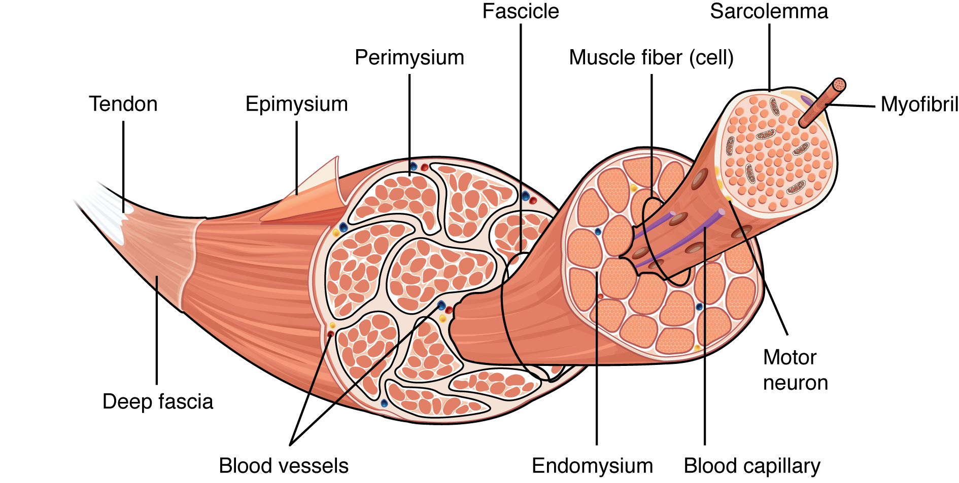
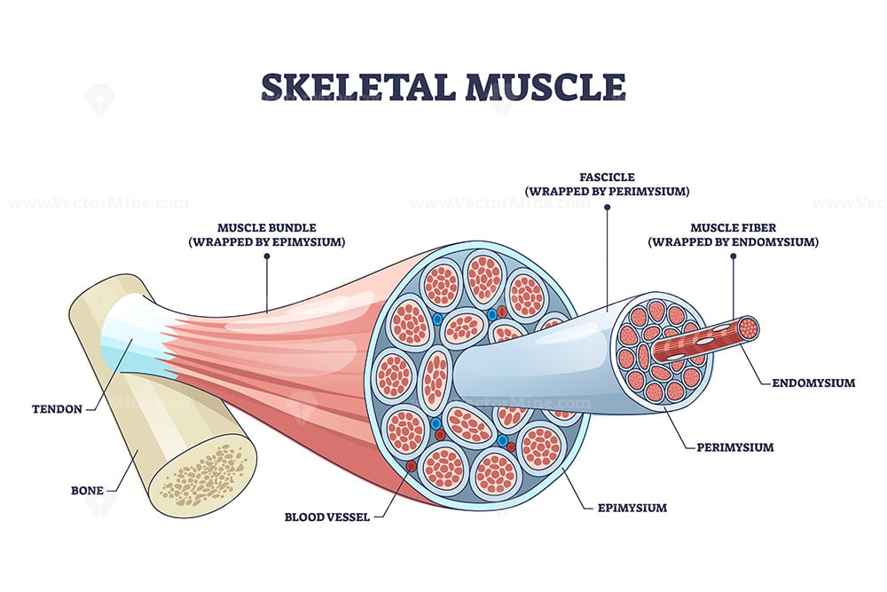
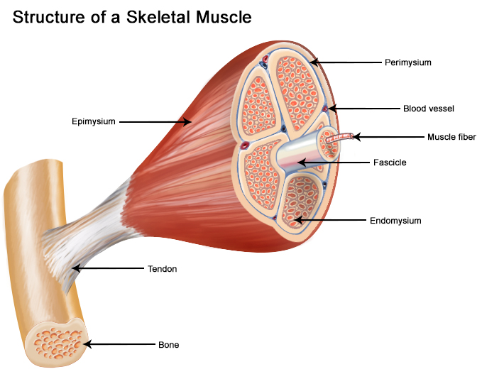
2. Gross Structure (Visible Structure – What You See Without Microscope)
A skeletal muscle is made of layers of connective tissue that hold everything together.
- Epimysium: The thick outer layer of connective tissue that covers the whole muscle (like a jacket).
- Perimysium: Connective tissue that wraps groups of muscle fibers into bundles called fascicles.
- Endomysium: Thin connective tissue around each single muscle fiber (muscle cell).
- Fascicle: A small bundle of 10–100 muscle fibers wrapped by perimysium.
- Muscle fiber (also called muscle cell): The long, cylinder-shaped cell that actually contracts. It runs the full length of the muscle.
- Tendon: Strong cord that connects muscle to bone (epimysium continues into tendon).
Think of it like this: Whole muscle = big rope → fascicles = smaller ropes inside → muscle fibers = threads in those ropes.
Here are clear labeled diagrams of the gross structure (showing epimysium, perimysium, endomysium, fascicle, muscle fiber):
3. Ultrastructure (Microscopic Details – Inside the Muscle Fiber)
Now zoom in with microscope/electron microscope level.
- Sarcolemma: The cell membrane of the muscle fiber (it can carry electrical signals).
- Sarcoplasm: The cytoplasm (fluid) inside the muscle fiber, containing organelles.
- Myofibrils: Long, thin threads running parallel inside the muscle fiber. Each myofibril is made of repeating units called sarcomeres — these do the actual contracting.
- Sarcomere: The smallest contractile unit — from one Z-line to the next Z-line.
- Sarcoplasmic Reticulum (SR): Special network like endoplasmic reticulum that stores Ca²⁺ (calcium ions) — very important for starting contraction.
- T-tubules (Transverse tubules): Small tubes from sarcolemma that go deep inside the fiber to carry electrical signals quickly.
- Triad: One T-tubule + two parts of SR (terminal cisternae) — this combo helps release calcium fast.
Here are detailed diagrams of muscle fiber ultrastructure (showing sarcolemma, myofibrils, SR, T-tubules, triad):
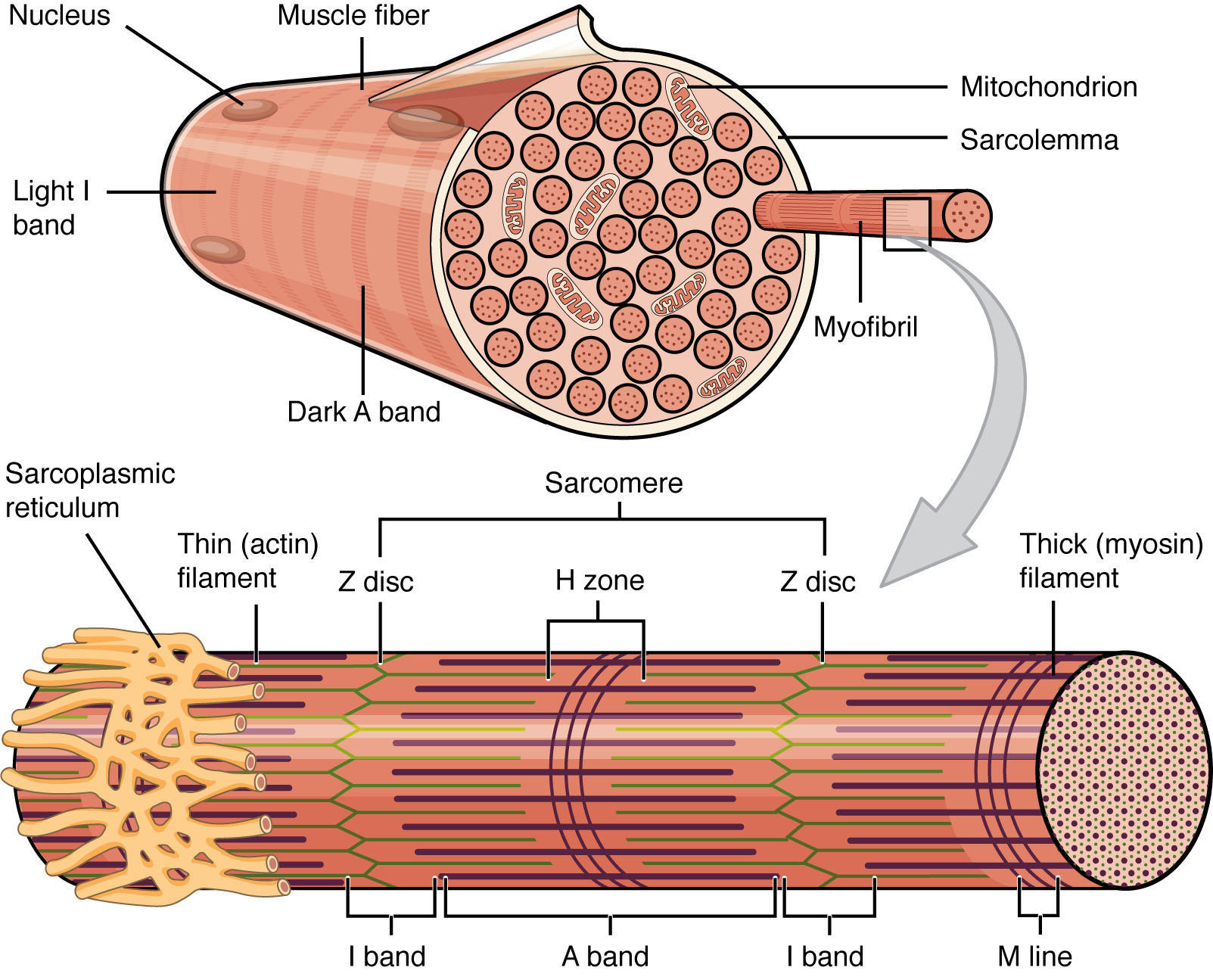
Sarcomere Bands and Zones (Very Important!)
- Z-line / Z-disk: Dark lines where thin filaments are attached (ends of sarcomere).
- A-band (Anisotropic band – dark under microscope): Full length of thick filaments — stays the same length during contraction.
- I-band (Isotropic band – light under microscope): Only thin filaments — shortens during contraction.
- H-zone / H-band (Hensen's zone): Center of A-band with only thick filaments (no thin overlap in relaxed state) — shortens or disappears during contraction.
- M-line / M-disk: Center line that holds thick filaments together.
Here are labeled sarcomere diagrams (relaxed state with bands):


Relaxed vs Contracted Sarcomere (Proof of Sliding)
In relaxed muscle: I-band and H-zone are wide. In contracted muscle: I-band and H-zone shorten (or H-zone disappears), Z-lines get closer, but A-band stays constant.
Diagrams showing relaxed vs contracted changes:


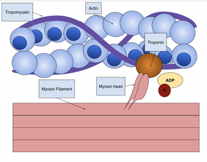
Thick and Thin Filaments
- Thick filaments: Made of myosin (has heads for pulling).
- Thin filaments: Made of actin (with tropomyosin covering sites + troponin that moves when calcium binds).
Diagram of thick (myosin) and thin (actin) filaments with tropomyosin and troponin:


.jpg)


+by+Blacklotus+Landscaping.jpg)


0 Comments